Show PDB file:   
         Plain Text   HTML   (compressed file size)
QuickSearch:   
by PDB,NDB,UniProt,PROSITE Code or Search Term(s)  
(-)Asym./Biol. Unit
(-)Asym./Biol. Unit - sites
collapse expand < >
Image Asym./Biol. Unit
Asym./Biol. Unit  (Jmol Viewer)
Image Asym./Biol. Unit - sites
Asym./Biol. Unit - sites  (Jmol Viewer)

(-) Description

Title :  THE HIGH RESOLUTION CRYSTAL STRUCTURE OF ROHEDSAIN, THE MAJOR CATHEPSIN L PROTEASE FROM T. BRUCEI RHODESIENSE, BOUND TO INHIBITOR K11002
 
Authors :  L. S. Brinen, R. Marion
Date :  21 Mar 07  (Deposition) - 01 Apr 08  (Release) - 24 Feb 09  (Revision)
Method :  X-RAY DIFFRACTION
Resolution :  1.16
Chains :  Asym./Biol. Unit :  A
Keywords :  Cysteine Protease, Trypanosoma Brucei, Neglected Disease, Hydrolase (Keyword Search: [Gene Ontology, PubMed, Web (Google))
 
Reference :  R. Marion, E. Hansell, C. Caffrey, W. R. Roush, L. S. Brinen
The High Resolution Structure Of Rhodesain, The Major Cathepsin L Protease From Trypanosoma Brucei Rhodesiense, Illustrates The Basis For Differences In Inhibition Profiles From Other Papain Family Cysteine Proteases
To Be Published
PubMed: search
(for further references see the PDB file header)

(-) Compounds

Molecule 1 - CYSTEINE PROTEASE
    ChainsA
    EC Number3.4.22.15
    EngineeredYES
    Expression SystemPICHIA PASTORIS
    Expression System PlasmidPPIC Z
    Expression System Taxid4922
    Expression System Vector TypePLASMID
    FragmentCATALYTIC DOMAIN
    GeneRHODESAIN
    MutationYES
    Organism ScientificTRYPANOSOMA BRUCEI RHODESIENSE
    Organism Taxid31286
    StrainRHODESIENSE

 Structural Features

(-) Chains, Units

  1
Asymmetric/Biological Unit A

Summary Information (see also Sequences/Alignments below)

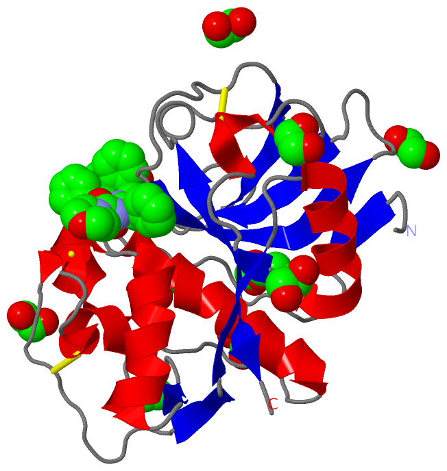
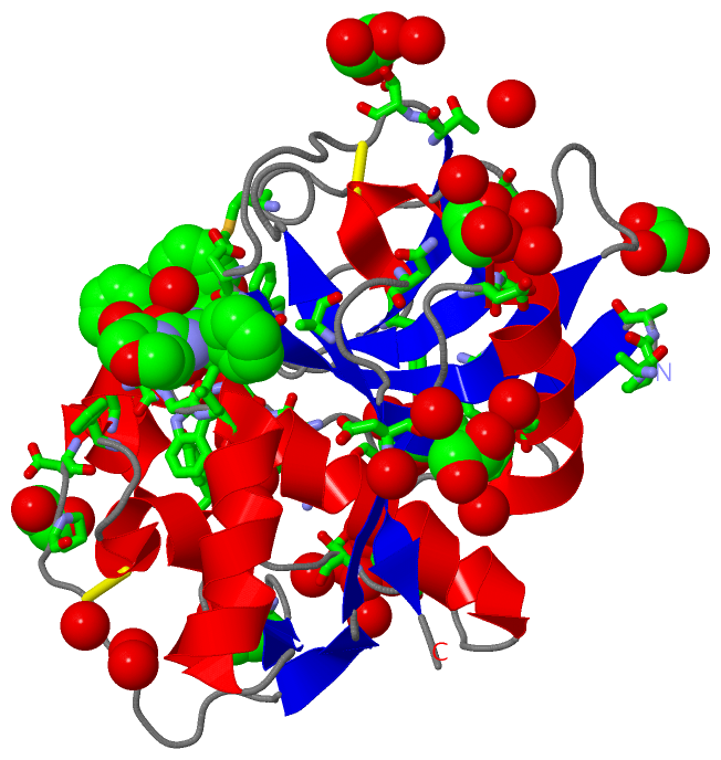
(-) Ligands, Modified Residues, Ions  (2, 12)

Asymmetric/Biological Unit (2, 12)
No.NameCountTypeFull Name
1EDO11Ligand/Ion1,2-ETHANEDIOL
2VS11Ligand/Ion3-[[N-[MORPHOLIN-N-YL]-CARBONYL]-PHENYLALANINYL-AMINO]-5- PHENYL-PENTANE-1-SULFONYLBENZENE

(-) Sites  (12, 12)

Asymmetric Unit (12, 12)
No.NameEvidenceResiduesDescription
01AC1SOFTWAREALA A:1 , PRO A:2 , ALA A:3 , GLN A:19 , GLY A:23 , CYS A:25 , TRP A:26 , PHE A:61 , CYS A:63 , GLY A:65 , GLY A:66 , LEU A:67 , ALA A:138 , MET A:145 , ASP A:161 , HIS A:162 , TRP A:184 , HOH A:440 , HOH A:607 , HOH A:691BINDING SITE FOR RESIDUE VS1 A 300
02AC2SOFTWAREASP A:121 , GLU A:122 , GLY A:199 , ASN A:206 , HOH A:510 , HOH A:558 , HOH A:561 , HOH A:683BINDING SITE FOR RESIDUE EDO A 901
03AC3SOFTWARELYS A:17 , ASP A:18 , TYR A:91 , HOH A:505 , HOH A:827BINDING SITE FOR RESIDUE EDO A 903
04AC4SOFTWARETHR A:14 , LEU A:45 , SER A:47 , HOH A:466 , HOH A:739 , HOH A:763BINDING SITE FOR RESIDUE EDO A 905
05AC5SOFTWAREASP A:18 , GLN A:19 , GLY A:20 , TRP A:184 , HOH A:607 , HOH A:627 , HOH A:665 , HOH A:826BINDING SITE FOR RESIDUE EDO A 906
06AC6SOFTWAREGLU A:97 , PRO A:99 , HOH A:609 , HOH A:624 , HOH A:782BINDING SITE FOR RESIDUE EDO A 907
07AC7SOFTWAREALA A:87 , THR A:153 , HOH A:494 , HOH A:653 , HOH A:713 , HOH A:808BINDING SITE FOR RESIDUE EDO A 908
08AC8SOFTWAREPRO A:15 , ASP A:117 , HOH A:424 , HOH A:445 , HOH A:513BINDING SITE FOR RESIDUE EDO A 910
09AC9SOFTWAREARG A:8 , THR A:14 , PRO A:15 , TYR A:193BINDING SITE FOR RESIDUE EDO A 911
10BC1SOFTWAREASP A:117 , TYR A:128 , HOH A:529 , HOH A:776BINDING SITE FOR RESIDUE EDO A 912
11BC2SOFTWARESER A:154 , HOH A:491 , HOH A:518 , HOH A:562 , HOH A:713BINDING SITE FOR RESIDUE EDO A 913
12BC3SOFTWAREPHE A:61 , HOH A:784BINDING SITE FOR RESIDUE EDO A 914

(-) SS Bonds  (3, 3)

Asymmetric/Biological Unit
No.Residues
1A:22 -A:63
2A:56 -A:101
3A:155 -A:203

(-) Cis Peptide Bonds  (1, 1)

Asymmetric/Biological Unit
No.Residues
1Asn A:174 -Pro A:175

 Sequence-Structure Mapping

(-) SAPs(SNPs)/Variants  (0, 0)

(no "SAP(SNP)/Variant" information available for 2P86)

(-) PROSITE Motifs  (0, 0)

(no "PROSITE Motif" information available for 2P86)

(-) Exons   (0, 0)

(no "Exon" information available for 2P86)

(-) Sequences/Alignments

Asymmetric/Biological Unit
   Reformat: Number of residues per line =  ('0' or empty: single-line sequence representation)
  Number of residues per labelling interval =   
  UniProt sequence: complete  aligned part    
   Show mapping: SCOP domains CATH domains Pfam domains Secondary structure (by author)
SAPs(SNPs) PROSITE motifs Exons
(details for a mapped element are shown in a popup box when the mouse pointer rests over it)
Chain A from PDB  Type:PROTEIN  Length:215
 aligned with Q95PM0_TRYBR | Q95PM0 from UniProtKB/TrEMBL  Length:450

    Alignment length:215
                                   135       145       155       165       175       185       195       205       215       225       235       245       255       265       275       285       295       305       315       325       335     
         Q95PM0_TRYBR   126 APAAVDWREKGAVTPVKDQGQCGSCWAFSTIGNIEGQWQVAGNPLVSLSEQMLVSCDTIDFGCGGGLMDNAFNWIVNSNGGNVFTEASYPYVSGNGEQPQCQMNGHEIGAAITDHVDLPQDEDAIAAYLAENGPLAIAVDATSFMDYNGGILTSCTSEQLDHGVLLVGYNDSSNPPYWIIKNSWSNMWGEDGYIRIEKGTNQCLMNQAVSSAVVG 340
               SCOP domains d2p86a_ A: automated matches                                                                                                                                                                                            SCOP domains
               CATH domains 2p86A00 A:1-215 Cysteine proteinases                                                                                                                                                                                    CATH domains
           Pfam domains (1) Peptidase_C1-2p86A01 A:1-213                                                                                                                                                                                         -- Pfam domains (1)
           Pfam domains (2) --------------------------------------------------------------------------------------------------------------------------------------------------------------------------------------------------------------------DUF Pfam domains (2)
         Sec.struct. author ...eee.................hhhhhhhhhhhhhhhhhh.......hhhhhhhhh...hhhhh.hhhhhhhhhhhhh..eee.......hhhhh...........eeee..eeee...hhhhhhhhhhhhh.eeeee...........ee.........eeeeeeeee......eeeee...........eeeee...hhhhh....eeee.. Sec.struct. author
                 SAPs(SNPs) ----------------------------------------------------------------------------------------------------------------------------------------------------------------------------------------------------------------------- SAPs(SNPs)
                    PROSITE ----------------------------------------------------------------------------------------------------------------------------------------------------------------------------------------------------------------------- PROSITE
                 Transcript ----------------------------------------------------------------------------------------------------------------------------------------------------------------------------------------------------------------------- Transcript
                 2p86 A   1 APAAVDWREKGAVTPVKDQGQCGSCWAFSTIGNIEGQWQVAGNPLVSLSEQMLVSCDTIDFGCGGGLMDNAFNWIVNSNGGNVFTEASYPYVSGNGEQPQCQMNGHEIGAAITDHVDLPQDEDAIAAYLAENGPLAIAVDATSFMDYNGGILTSCTSEQLDHGVLLVGYNDASNPPYWIIKNSWSNMWGEDGYIRIEKGTNQCLMNQAVSSAVVG 215
                                    10        20        30        40        50        60        70        80        90       100       110       120       130       140       150       160       170       180       190       200       210     

   Legend:   → Mismatch (orange background)
  - → Gap (green background, '-', border residues have a numbering label)
    → Modified Residue (blue background, lower-case, 'x' indicates undefined single-letter code, labelled with number + name)
  x → Chemical Group (purple background, 'x', labelled with number + name, e.g. ACE or NH2)
  extra numbering lines below/above indicate numbering irregularities and modified residue names etc., number ends below/above '|'

 Classification and Annotation

(-) SCOP Domains  (1, 1)

Asymmetric/Biological Unit

(-) CATH Domains  (1, 1)

Asymmetric/Biological Unit
(-)
Class: Alpha Beta (26913)

(-) Pfam Domains  (2, 2)

Asymmetric/Biological Unit

(-) Gene Ontology  (5, 5)

Asymmetric/Biological Unit(hide GO term definitions)
Chain A   (Q95PM0_TRYBR | Q95PM0)
molecular function
    GO:0004197    cysteine-type endopeptidase activity    Catalysis of the hydrolysis of internal, alpha-peptide bonds in a polypeptide chain by a mechanism in which the sulfhydryl group of a cysteine residue at the active center acts as a nucleophile.
    GO:0008234    cysteine-type peptidase activity    Catalysis of the hydrolysis of peptide bonds in a polypeptide chain by a mechanism in which the sulfhydryl group of a cysteine residue at the active center acts as a nucleophile.
    GO:0016787    hydrolase activity    Catalysis of the hydrolysis of various bonds, e.g. C-O, C-N, C-C, phosphoric anhydride bonds, etc. Hydrolase is the systematic name for any enzyme of EC class 3.
    GO:0008233    peptidase activity    Catalysis of the hydrolysis of a peptide bond. A peptide bond is a covalent bond formed when the carbon atom from the carboxyl group of one amino acid shares electrons with the nitrogen atom from the amino group of a second amino acid.
biological process
    GO:0006508    proteolysis    The hydrolysis of proteins into smaller polypeptides and/or amino acids by cleavage of their peptide bonds.

 Visualization

(-) Interactive Views

Asymmetric/Biological Unit
  Complete Structure
    Jena3D(integrated viewing of ligand, site, SAP, PROSITE, SCOP information)
    WebMol | AstexViewer[tm]@PDBe
(Java Applets, require no local installation except for Java; loading may be slow)
    STRAP
(Java WebStart application, automatic local installation, requires Java; full application with system access!)
    RasMol
(require local installation)
    Molscript (VRML)
(requires installation of a VRML viewer; select preferred view via VRML and generate a mono or stereo PDF format file)
 
  Ligands, Modified Residues, Ions
    EDO  [ RasMol | Jena3D ]  +environment [ RasMol | Jena3D ]
    VS1  [ RasMol | Jena3D ]  +environment [ RasMol | Jena3D ]
 
  Sites
    AC1  [ RasMol ]  +environment [ RasMol ]
    AC2  [ RasMol ]  +environment [ RasMol ]
    AC3  [ RasMol ]  +environment [ RasMol ]
    AC4  [ RasMol ]  +environment [ RasMol ]
    AC5  [ RasMol ]  +environment [ RasMol ]
    AC6  [ RasMol ]  +environment [ RasMol ]
    AC7  [ RasMol ]  +environment [ RasMol ]
    AC8  [ RasMol ]  +environment [ RasMol ]
    AC9  [ RasMol ]  +environment [ RasMol ]
    BC1  [ RasMol ]  +environment [ RasMol ]
    BC2  [ RasMol ]  +environment [ RasMol ]
    BC3  [ RasMol ]  +environment [ RasMol ]
 
  Cis Peptide Bonds
    Asn A:174 - Pro A:175   [ RasMol ]  
 

(-) Still Images

Jmol
  protein: cartoon or spacefill or dots and stick; nucleic acid: cartoon and stick; ligands: spacefill; active site: stick
Molscript
  protein, nucleic acid: cartoon; ligands: spacefill; active site: ball and stick

 Databases and Analysis Tools

(-) Databases

Access by PDB/NDB ID
  2p86
    Family and Domain InformationProDom | SYSTERS
    General Structural InformationGlycoscienceDB | MMDB | NDB | OCA | PDB | PDBe | PDBj | PDBsum | PDBWiki | PQS | PROTEOPEDIA
    Orientation in MembranesOPM
    Protein SurfaceSURFACE
    Secondary StructureDSSP (structure derived) | HSSP (homology derived)
    Structural GenomicsGeneCensus
    Structural NeighboursCE | VAST
    Structure ClassificationCATH | Dali | SCOP
    Validation and Original DataBMRB Data View | BMRB Restraints Grid | EDS | PROCHECK | RECOORD | WHAT_CHECK
 
Access by UniProt ID/Accession number
  Q95PM0_TRYBR | Q95PM0
    Comparative Protein Structure ModelsModBase
    Genomic InformationEnsembl
    Protein-protein InteractionDIP
    Sequence, Family and Domain InformationInterPro | Pfam | SMART | UniProtKB/TrEMBL
 
Access by Enzyme Classificator   (EC Number)
  3.4.22.15
    General Enzyme InformationBRENDA | EC-PDB | Enzyme | IntEnz
    PathwayKEGG | MetaCyc
 
Access by Disease Identifier   (MIM ID)
  (no 'MIM ID' available)
    Disease InformationOMIM
 
Access by GenAge ID
  (no 'GenAge ID' available)
    Age Related InformationGenAge

(-) Analysis Tools

Access by PDB/NDB ID
    Domain InformationXDom
    Interatomic Contacts of Structural UnitsCSU
    Ligand-protein ContactsLPC
    Protein CavitiescastP
    Sequence and Secondary StructurePDBCartoon
    Structure AlignmentSTRAP(Java WebStart application, automatic local installation, requires Java; full application with system access!)
    Structure and Sequence BrowserSTING
 
Access by UniProt ID/Accession number
  Q95PM0_TRYBR | Q95PM0
    Protein Disorder PredictionDisEMBL | FoldIndex | GLOBPLOT (for more information see DisProt)

 Related Entries

(-) Entries Sharing at Least One Protein Chain (UniProt ID)

UniProtKB/TrEMBL
        Q95PM0_TRYBR | Q95PM02p7u

(-) Related Entries Specified in the PDB File

2p7u THE CRYSTAL STRUCTURE OF RHODESAIN BOUND TO INHIBITOR K11777