Show PDB file:   
         Plain Text   HTML   (compressed file size)
QuickSearch:   
by PDB,NDB,UniProt,PROSITE Code or Search Term(s)  
(-)Asym./Biol. Unit
(-)Asym./Biol. Unit - sites
collapse expand < >
Image Asym./Biol. Unit
Asym./Biol. Unit  (Jmol Viewer)
Image Asym./Biol. Unit - sites
Asym./Biol. Unit - sites  (Jmol Viewer)

(-) Description

Title :  CRYSTAL STRUCTURE OF BORDETELLA PERTUSSIS HOLO FERRIC BINDING PROTEIN BOUND WITH TWO SYNERGISTIC OXALATE ANIONS
 
Authors :  S. A. L. Tom-Yew, M. E. P. Murphy
Date :  16 Feb 07  (Deposition) - 26 Feb 08  (Release) - 24 Feb 09  (Revision)
Method :  X-RAY DIFFRACTION
Resolution :  1.49
Chains :  Asym./Biol. Unit :  A
Keywords :  Alpha-Beta-Alpha Units, Anti-Parallel Beta-Strands Hinge Region, Metal Binding Protein (Keyword Search: [Gene Ontology, PubMed, Web (Google))
 
Reference :  S. A. L. Tom-Yew, B. H. Shilton, E. G. Bekker, E. I. Tocheva, M. E. P. Murphy
Anion-Dependent Hinge Motion In Ferric Binding Proteins
To Be Published
PubMed: search
(for further references see the PDB file header)

(-) Compounds

Molecule 1 - PUTATIVE IRON BINDING PROTEIN
    ChainsA
    EngineeredYES
    Expression SystemESCHERICHIA COLI BL21(DE3)
    Expression System PlasmidPET28A
    Expression System StrainBL21 (DE3)
    Expression System Taxid469008
    Expression System Vector TypePLASMID
    GeneBFBPA
    Organism ScientificBORDETELLA PERTUSSIS TOHAMA I
    Organism Taxid257313
    StrainTOHAMA I

 Structural Features

(-) Chains, Units

  1
Asymmetric/Biological Unit A

Summary Information (see also Sequences/Alignments below)
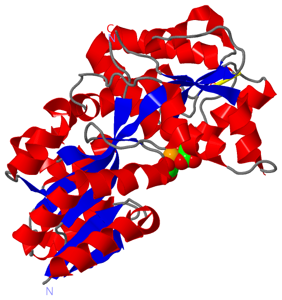
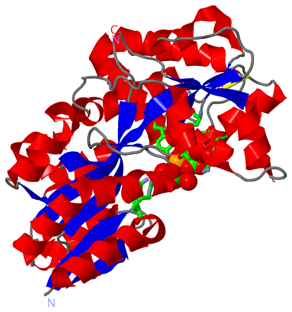
(-) Ligands, Modified Residues, Ions  (2, 3)

Asymmetric/Biological Unit (2, 3)
No.NameCountTypeFull Name
1FE1Ligand/IonFE (III) ION
2OXL2Ligand/IonOXALATE ION

(-) Sites  (3, 3)

Asymmetric Unit (3, 3)
No.NameEvidenceResiduesDescription
1AC1SOFTWARETYR A:143 , TYR A:200BINDING SITE FOR RESIDUE FE A 324
2AC2SOFTWAREARG A:137 , TYR A:143 , GLY A:178 , ASP A:179 , ARG A:180 , TYR A:200 , HOH A:339 , HOH A:519BINDING SITE FOR RESIDUE OXL A 325
3AC3SOFTWAREARG A:14 , TYR A:143 , TYR A:200 , HOH A:384 , HOH A:501 , HOH A:546BINDING SITE FOR RESIDUE OXL A 326

(-) SS Bonds  (1, 1)

Asymmetric/Biological Unit
No.Residues
1A:135 -A:191

(-) Cis Peptide Bonds  (0, 0)

(no "Cis Peptide Bond" information available for 2OWS)

 Sequence-Structure Mapping

(-) SAPs(SNPs)/Variants  (0, 0)

(no "SAP(SNP)/Variant" information available for 2OWS)

(-) PROSITE Motifs  (0, 0)

(no "PROSITE Motif" information available for 2OWS)

(-) Exons   (0, 0)

(no "Exon" information available for 2OWS)

(-) Sequences/Alignments

Asymmetric/Biological Unit
   Reformat: Number of residues per line =  ('0' or empty: single-line sequence representation)
  Number of residues per labelling interval =   
  UniProt sequence: complete  aligned part    
   Show mapping: SCOP domains CATH domains Pfam domains Secondary structure (by author)
SAPs(SNPs) PROSITE motifs Exons
(details for a mapped element are shown in a popup box when the mouse pointer rests over it)
Chain A from PDB  Type:PROTEIN  Length:314
 aligned with Q7VXW9_BORPE | Q7VXW9 from UniProtKB/TrEMBL  Length:349

    Alignment length:318
                                    41        51        61        71        81        91       101       111       121       131       141       151       161       171       181       191       201       211       221       231       241       251       261       271       281       291       301       311       321       331       341        
         Q7VXW9_BORPE    32 DEVSLYTTREPKLIQPLLDAFAKDSGIKVNTVFVKDGLLERVRAEGDKSPADVLMTVDIGNLIDLVNGGVTQKIQSQTLDSVVPANLRGAEGSWYALSLRDRVLYVEKDLKLDSFRYGDLADPKWKGKVCIRSGQHPYNTALVAAMIAHDGAEATEKWLRGVKANLARKAAGGDRDVARDILGGICDIGLANAYYVGHMKNAEPGTDARKWGDAIKVVRPTFATAKDGGTHVNISGAAVAAHAPNKANAVKLLEYLVSEPAQTLYAQANYEYPVRAGVKLDAVVASFGPLKVDTLPVAEIAKYRKQASELVDKVGFDN 349
               SCOP domains d2owsa_ A: automated matches                                                                                                                                                                                                                                                                                                   SCOP domains
               CATH domains 2owsA01 A:6-102,A:234-283 Periplasmic binding protein-like II                                    2owsA02 A:103-233,A:284-323 Periplasmic binding protein-like II                                                                    2owsA01 A:6-102,A:234-283                         2owsA02 A:103-233,A:284-323              CATH domains
               Pfam domains -----------------------------------------------SBP_bac_6-2owsA01 A:53-298                                                                                                                                                                                                                            ------------------------- Pfam domains
         Sec.struct. author .eeeeee..hhhhhhhhhhhhhhhhh.eeeeee...hhhhhhhhhhhhh...eeeeehhhhhhhhhhh..ee...hhhhhhhhhhhhhhhhhh.eeeeeeeeeeeee........hhhhhhhhhhh..ee.....hhhhhhhhhhhhhhhhhhhhhhhhhhhhhhh.......hhhhhhhhhh....eeeeehhhhhhhhh....hhhhhhhhheeee.....----....eeeeeeeee....hhhhhhhhhhhhhhhhhhhhhhhhh..ee.......hhhhhhh.........hhhhhhhhhhhhhhhhhh.... Sec.struct. author
                 SAPs(SNPs) ------------------------------------------------------------------------------------------------------------------------------------------------------------------------------------------------------------------------------------------------------------------------------------------------------------------------------ SAPs(SNPs)
                    PROSITE ------------------------------------------------------------------------------------------------------------------------------------------------------------------------------------------------------------------------------------------------------------------------------------------------------------------------------ PROSITE
                 Transcript ------------------------------------------------------------------------------------------------------------------------------------------------------------------------------------------------------------------------------------------------------------------------------------------------------------------------------ Transcript
                 2ows A   6 DEVSLYTTREPKLIQPLLDAFAKDSGIKVNTVFVKDGLLERVRAEGDKSPADVLMTVDIGNLIDLVNGGVTQKIQSQTLDSVVPANLRGAEGSWYALSLRDRVLYVEKDLKLDSFRYGDLADPKWKGKVCIRSGQHPYNTALVAAMIAHDGAEATEKWLRGVKANLARKAAGGDRDVARDILGGICDIGLANAYYVGHMKNAEPGTDARKWGDAIKVVRPTFA----GGTHVNISGAAVAAHAPNKANAVKLLEYLVSEPAQTLYAQANYEYPVRAGVKLDAVVASFGPLKVDTLPVAEIAKYRKQASELVDKVGFDN 323
                                    15        25        35        45        55        65        75        85        95       105       115       125       135       145       155       165       175       185       195       205       215       225  |    235       245       255       265       275       285       295       305       315        
                                                                                                                                                                                                                                                        228  233                                                                                          

   Legend:   → Mismatch (orange background)
  - → Gap (green background, '-', border residues have a numbering label)
    → Modified Residue (blue background, lower-case, 'x' indicates undefined single-letter code, labelled with number + name)
  x → Chemical Group (purple background, 'x', labelled with number + name, e.g. ACE or NH2)
  extra numbering lines below/above indicate numbering irregularities and modified residue names etc., number ends below/above '|'

 Classification and Annotation

(-) SCOP Domains  (1, 1)

Asymmetric/Biological Unit

(-) CATH Domains  (1, 2)

Asymmetric/Biological Unit
(-)
Class: Alpha Beta (26913)

(-) Pfam Domains  (1, 1)

Asymmetric/Biological Unit
(-)
Clan: PBP (391)

(-) Gene Ontology  (1, 1)

Asymmetric/Biological Unit(hide GO term definitions)
Chain A   (Q7VXW9_BORPE | Q7VXW9)
molecular function
    GO:0046872    metal ion binding    Interacting selectively and non-covalently with any metal ion.

 Visualization

(-) Interactive Views

Asymmetric/Biological Unit
  Complete Structure
    Jena3D(integrated viewing of ligand, site, SAP, PROSITE, SCOP information)
    WebMol | AstexViewer[tm]@PDBe
(Java Applets, require no local installation except for Java; loading may be slow)
    STRAP
(Java WebStart application, automatic local installation, requires Java; full application with system access!)
    RasMol
(require local installation)
    Molscript (VRML)
(requires installation of a VRML viewer; select preferred view via VRML and generate a mono or stereo PDF format file)
 
  Ligands, Modified Residues, Ions
    FE  [ RasMol | Jena3D ]  +environment [ RasMol | Jena3D ]
    OXL  [ RasMol | Jena3D ]  +environment [ RasMol | Jena3D ]
 
  Sites
    AC1  [ RasMol ]  +environment [ RasMol ]
    AC2  [ RasMol ]  +environment [ RasMol ]
    AC3  [ RasMol ]  +environment [ RasMol ]
 
  Cis Peptide Bonds
(no "Cis Peptide Bonds" information available for 2ows)
 

(-) Still Images

Jmol
  protein: cartoon or spacefill or dots and stick; nucleic acid: cartoon and stick; ligands: spacefill; active site: stick
Molscript
  protein, nucleic acid: cartoon; ligands: spacefill; active site: ball and stick

 Databases and Analysis Tools

(-) Databases

Access by PDB/NDB ID
  2ows
    Family and Domain InformationProDom | SYSTERS
    General Structural InformationGlycoscienceDB | MMDB | NDB | OCA | PDB | PDBe | PDBj | PDBsum | PDBWiki | PQS | PROTEOPEDIA
    Orientation in MembranesOPM
    Protein SurfaceSURFACE
    Secondary StructureDSSP (structure derived) | HSSP (homology derived)
    Structural GenomicsGeneCensus
    Structural NeighboursCE | VAST
    Structure ClassificationCATH | Dali | SCOP
    Validation and Original DataBMRB Data View | BMRB Restraints Grid | EDS | PROCHECK | RECOORD | WHAT_CHECK
 
Access by UniProt ID/Accession number
  Q7VXW9_BORPE | Q7VXW9
    Comparative Protein Structure ModelsModBase
    Genomic InformationEnsembl
    Protein-protein InteractionDIP
    Sequence, Family and Domain InformationInterPro | Pfam | SMART | UniProtKB/TrEMBL
 
Access by Enzyme Classificator   (EC Number)
  (no 'Enzyme Classificator' available)
    General Enzyme InformationBRENDA | EC-PDB | Enzyme | IntEnz
    PathwayKEGG | MetaCyc
 
Access by Disease Identifier   (MIM ID)
  (no 'MIM ID' available)
    Disease InformationOMIM
 
Access by GenAge ID
  (no 'GenAge ID' available)
    Age Related InformationGenAge

(-) Analysis Tools

Access by PDB/NDB ID
    Domain InformationXDom
    Interatomic Contacts of Structural UnitsCSU
    Ligand-protein ContactsLPC
    Protein CavitiescastP
    Sequence and Secondary StructurePDBCartoon
    Structure AlignmentSTRAP(Java WebStart application, automatic local installation, requires Java; full application with system access!)
    Structure and Sequence BrowserSTING
 
Access by UniProt ID/Accession number
  Q7VXW9_BORPE | Q7VXW9
    Protein Disorder PredictionDisEMBL | FoldIndex | GLOBPLOT (for more information see DisProt)

 Related Entries

(-) Entries Sharing at Least One Protein Chain (UniProt ID)

UniProtKB/TrEMBL
        Q7VXW9_BORPE | Q7VXW91y9u 2owt

(-) Related Entries Specified in the PDB File

1y9u APO FORM OF SAME PROTEIN