|
|
|
|
 Description
|
|
Compounds
|
||||||||||||||||||||||||||||||||||||||||||||||||||||
Chains, Units
Summary Information (see also Sequences/Alignments below) |
Ligands, Modified Residues, Ions (1, 1)
Asymmetric/Biological Unit (1, 1)
|
Sites (1, 1)
Asymmetric Unit (1, 1)
|
SS Bonds (0, 0)| (no "SS Bond" information available for 2NT4) |
Cis Peptide Bonds (1, 1)
Asymmetric/Biological Unit
|
||||||||
SAPs(SNPs)/Variants (0, 0)| (no "SAP(SNP)/Variant" information available for 2NT4) |
PROSITE Motifs (0, 0)| (no "PROSITE Motif" information available for 2NT4) |
Exons (0, 0)| (no "Exon" information available for 2NT4) |
Sequences/Alignments
Asymmetric/Biological UnitChain A from PDB Type:PROTEIN Length:126 aligned with O68522_MYXXA | O68522 from UniProtKB/TrEMBL Length:562 Alignment length:126 1 | 8 18 28 38 48 58 68 78 88 98 108 118 O68522_MYXXA - --MSKKILIVESDTALSATLRSALEGRGFTVDETTDGKGSVEQIRRDRPDLVVLAVDLSAGQNGYLICGKLKKDDDLKNVPIVIIGNPDGFAQHRKLKAHADEYVAKPVDADQLVERAGALIGFPE 124 SCOP domains d2nt4a_ A: automated matches SCOP domains CATH domains ------------------------------------------------------------------------------------------------------------------------------ CATH domains Pfam domains ------Response_reg-2nt4A01 A:5-116 -------- Pfam domains SAPs(SNPs) ------------------------------------------------------------------------------------------------------------------------------ SAPs(SNPs) PROSITE ------------------------------------------------------------------------------------------------------------------------------ PROSITE Transcript ------------------------------------------------------------------------------------------------------------------------------ Transcript 2nt4 A -1 SHMSKKILIVESDTALSATLRSALEGRGFTVDETTDGKGSVEQIRRDRPDLVVLAVDLSAGQNGYLICGKLKKDDDLKNVPIVIIGNPDGFAQFRKLKAHADEYVAKPVDADQLVERAGALIGFPE 124 8 18 28 38 48 58 68 78 88 98 108 118
|
||||||||||||||||||||
SCOP Domains (1, 1)
Asymmetric/Biological Unit
 |
CATH Domains (0, 0)| (no "CATH Domain" information available for 2NT4) |
Pfam Domains (1, 1)
Asymmetric/Biological Unit
|
Gene Ontology (2, 2)|
Asymmetric/Biological Unit(hide GO term definitions) Chain A (O68522_MYXXA | O68522)
|
||||||||||||||||||||||||
Interactive Views
|
|||||||||||||||||||||||||||||||||||||||||||||||||||||||||||||||||||||||||||||||||||||||||||||||||||||||||||||||||||||||
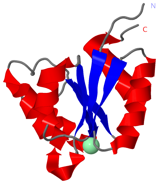
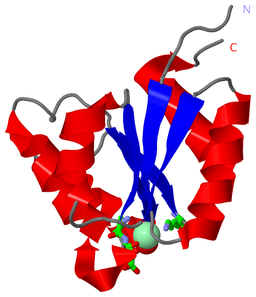
Still Images
|
||||||||||||||||
Databases
|
||||||||||||||||||||||||||||||||||||||||||||||||||||||||||||||||||||||||||||||||||||||||||||||||||||||||||||||||||||||||||||||||||||||||||||||||||||||||||||||||
Analysis Tools
|
|||||||||||||||||||||||||||||||||||||||||||||||||||||||||||||
Entries Sharing at Least One Protein Chain (UniProt ID)
Related Entries Specified in the PDB File
|
|