|
|
|
|
 Description
|
|
Compounds
|
||||||||||||||||||||||||||||||||||||||||||||||||||||||||||||||||||||||||||
Chains, Units
Summary Information (see also Sequences/Alignments below) |
Ligands, Modified Residues, Ions (0, 0)| (no "Ligand,Modified Residues,Ions" information available for 2N01) |
Sites (0, 0)| (no "Site" information available for 2N01) |
SS Bonds (0, 0)| (no "SS Bond" information available for 2N01) |
Cis Peptide Bonds (0, 0)| (no "Cis Peptide Bond" information available for 2N01) |
SAPs(SNPs)/Variants (0, 0)| (no "SAP(SNP)/Variant" information available for 2N01) |
PROSITE Motifs (0, 0)| (no "PROSITE Motif" information available for 2N01) |
Exons (0, 0)| (no "Exon" information available for 2N01) |
Sequences/Alignments
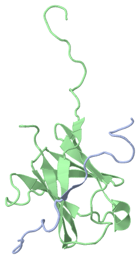
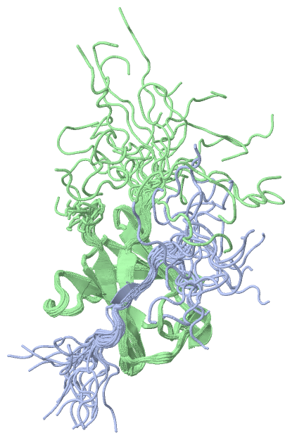
NMR Structure
Chain A from PDB Type:PROTEIN Length:23
SCOP domains ----------------------- SCOP domains
CATH domains ----------------------- CATH domains
Pfam domains ----------------------- Pfam domains
SAPs(SNPs) ----------------------- SAPs(SNPs)
PROSITE ----------------------- PROSITE
Transcript ----------------------- Transcript
2n01 A 24 TKPAPDFGGRWKHVNHFDEAPTE 46
33 43
Chain B from PDB Type:PROTEIN Length:106
SCOP domains ---------------------------------------------------------------------------------------------------------- SCOP domains
CATH domains ---------------------------------------------------------------------------------------------------------- CATH domains
Pfam domains ---------------------------------------------------------------------------------------------------------- Pfam domains
SAPs(SNPs) ---------------------------------------------------------------------------------------------------------- SAPs(SNPs)
PROSITE ---------------------------------------------------------------------------------------------------------- PROSITE
Transcript ---------------------------------------------------------------------------------------------------------- Transcript
2n01 B 150 GSHMNAKILKDRRYYYDYDYATRTKKSWLIPSRVYDDGKFTYINMDLTRFPTGNFPAVFAREKEHAEDFLVNTTVEGNTLIVHGTYPFLVVRHGDNVVGLRRNKQK 255
159 169 179 189 199 209 219 229 239 249
|
||||||||||||||||||||
SCOP Domains (0, 0)| (no "SCOP Domain" information available for 2N01) |
CATH Domains (0, 0)| (no "CATH Domain" information available for 2N01) |
Pfam Domains (0, 0)| (no "Pfam Domain" information available for 2N01) |
Gene Ontology (0, 0)|
NMR Structure(hide GO term definitions)
(no "Gene Ontology" information available for 2N01)
|
Interactive Views
|
||||||||||||||||||||||||||||||||||||||||||||||||||||||||||||||||||||||||||||||||||||||||||||||||||||||||||||||||||||
Still Images
|
||||||||||||||||
Databases
|
||||||||||||||||||||||||||||||||||||||||||||||||||||||||||||||||||||||||||||||||||||||||||||||||||||||||||||||||||||||||||||||||||||||||||||||||||||||||||||||||||||||||||||||||||||||||||
Analysis Tools
|
||||||||||||||||||||||||||||||||||||||||||||||||||||||||||||||||||||||||
Entries Sharing at Least One Protein Chain (UniProt ID)
Related Entries Specified in the PDB File
|
|