Show PDB file:   
         Plain Text   HTML   (compressed file size)
QuickSearch:   
by PDB,NDB,UniProt,PROSITE Code or Search Term(s)  
(-)NMR Structure - model 1
(-)NMR Structure - all models
collapse expand < >
Image NMR Structure - model 1
NMR Structure - model 1  (Jmol Viewer)
Image NMR Structure - all models
NMR Structure - all models  (Jmol Viewer)

(-) Description

Title :  STRUCTURE OF PLASMODIUM YOELII MEROZOITE SURFACE PROTEIN 1 - C-TERMINAL DOMAIN
 
Authors :  R. D. Curd, B. Birdsall, M. Kadekoppala, S. Ogun, G. Kelly, A. A. Holder
Date :  03 Nov 13  (Deposition) - 12 Feb 14  (Release) - 12 Feb 14  (Revision)
Method :  SOLUTION NMR
Resolution :  NOT APPLICABLE
Chains :  NMR Structure  :  A  (20x)
NMR Structure *:  A  (1x)
Keywords :  Membrane Protein (Keyword Search: [Gene Ontology, PubMed, Web (Google))
 
Reference :  R. D. Curd, B. Birdsall, M. Kadekoppala, S. A. Ogun, G. Kelly, A. A. Holder
The Structure Of Plasmodium Yoelii Merozoite Surface Protei 119, Antibody Specificity And Implications For Malaria Vaccine Design
Open Biology V. 4 30091 2014
PubMed-ID: 24403012  |  Reference-DOI: 10.1098/RSOB.130091

(-) Compounds

Molecule 1 - MEROZOITE SURFACE PROTEIN 1
    ChainsA
    EngineeredYES
    Expression SystemKOMAGATAELLA PASTORIS
    Expression System Taxid4922
    Expression System VectorPPIC9K
    Expression System Vector TypeVECTOR
    FragmentC-TERMINAL DOMAIN, UNP RESIDUES 1656-1754
    GeneMSP-1
    MutationYES
    Organism ScientificPLASMODIUM YOELII YOELII
    Organism Taxid73239

 Structural Features

(-) Chains, Units

  1
NMR Structure (20x)A
NMR Structure * (1x)A

Summary Information (see also Sequences/Alignments below)

(-) Ligands, Modified Residues, Ions  (0, 0)

(no "Ligand,Modified Residues,Ions" information available for 2MGP)

(-) Sites  (0, 0)

(no "Site" information available for 2MGP)
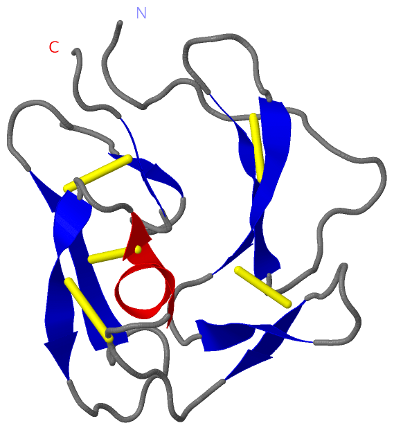
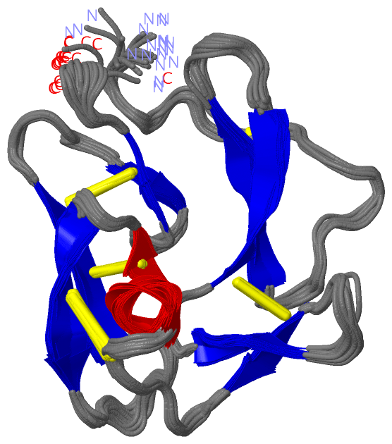
(-) SS Bonds  (5, 5)

NMR Structure
No.Residues
1A:8 -A:20
2A:32 -A:44
3A:52 -A:65
4A:59 -A:79
5A:81 -A:95

(-) Cis Peptide Bonds  (0, 0)

(no "Cis Peptide Bond" information available for 2MGP)

 Sequence-Structure Mapping

(-) SAPs(SNPs)/Variants  (0, 0)

(no "SAP(SNP)/Variant" information available for 2MGP)

(-) PROSITE Motifs  (0, 0)

(no "PROSITE Motif" information available for 2MGP)

(-) Exons   (0, 0)

(no "Exon" information available for 2MGP)

(-) Sequences/Alignments

NMR Structure
   Reformat: Number of residues per line =  ('0' or empty: single-line sequence representation)
  Number of residues per labelling interval =   
  UniProt sequence: complete  aligned part    
   Show mapping: SCOP domains CATH domains Pfam domains Secondary structure (by author)
SAPs(SNPs) PROSITE motifs Exons
(details for a mapped element are shown in a popup box when the mouse pointer rests over it)
Chain A from PDB  Type:PROTEIN  Length:99
 aligned with MSP1_PLAYO | P13828 from UniProtKB/Swiss-Prot  Length:1772

    Alignment length:99
                                  1665      1675      1685      1695      1705      1715      1725      1735      1745         
          MSP1_PLAYO   1656 GVDPKHVCVDTRDIPKNAGCFRDDNGTEEWRCLLGYKKGEGNTCVENNNPTCDINNGGCDPTASCQNAESTENSKKIICTCKEPTPNAYYEGVFCSSSS 1754
               SCOP domains d2mgpa1 A:1-48                                  d2mgpa2 A:49-99 Merozoite surface protein 1 (MSP-1) SCOP domains
               CATH domains --------------------------------------------------------------------------------------------------- CATH domains
               Pfam domains --------------------------------------------------------------------------------------------------- Pfam domains
         Sec.struct. author .................eeeee.....eeeee...eee.....eee.......hhhhhh...eeeee.........eeeee......ee....ee.... Sec.struct. author
                 SAPs(SNPs) --------------------------------------------------------------------------------------------------- SAPs(SNPs)
                    PROSITE --------------------------------------------------------------------------------------------------- PROSITE
                 Transcript --------------------------------------------------------------------------------------------------- Transcript
                2mgp A    1 GVDPKHVCVDTRDIPKNAGCFRDDDGTEEWRCLLGYKKGEGNTCVENNNPTCDINNGGCDPTASCQNAESTENSKKIICTCKEPTPNAYYEGVFCSSSS   99
                                    10        20        30        40        50        60        70        80        90         

   Legend:   → Mismatch (orange background)
  - → Gap (green background, '-', border residues have a numbering label)
    → Modified Residue (blue background, lower-case, 'x' indicates undefined single-letter code, labelled with number + name)
  x → Chemical Group (purple background, 'x', labelled with number + name, e.g. ACE or NH2)
  extra numbering lines below/above indicate numbering irregularities and modified residue names etc., number ends below/above '|'

 Classification and Annotation

(-) SCOP Domains  (1, 2)

NMR Structure

(-) CATH Domains  (0, 0)

(no "CATH Domain" information available for 2MGP)

(-) Pfam Domains  (0, 0)

(no "Pfam Domain" information available for 2MGP)

(-) Gene Ontology  (4, 4)

NMR Structure(hide GO term definitions)
Chain A   (MSP1_PLAYO | P13828)
biological process
    GO:0009405    pathogenesis    The set of specific processes that generate the ability of an organism to induce an abnormal, generally detrimental state in another organism.
cellular component
    GO:0031225    anchored component of membrane    The component of a membrane consisting of the gene products that are tethered to the membrane only by a covalently attached anchor, such as a lipid group that is embedded in the membrane. Gene products with peptide sequences that are embedded in the membrane are excluded from this grouping.
    GO:0016020    membrane    A lipid bilayer along with all the proteins and protein complexes embedded in it an attached to it.
    GO:0005886    plasma membrane    The membrane surrounding a cell that separates the cell from its external environment. It consists of a phospholipid bilayer and associated proteins.

 Visualization

(-) Interactive Views

NMR Structure
  Complete Structure
    Jena3D(integrated viewing of ligand, site, SAP, PROSITE, SCOP information)
    WebMol | AstexViewer[tm]@PDBe
(Java Applets, require no local installation except for Java; loading may be slow)
    STRAP
(Java WebStart application, automatic local installation, requires Java; full application with system access!)
    RasMol
(require local installation)
    Molscript (VRML)
(requires installation of a VRML viewer; select preferred view via VRML and generate a mono or stereo PDF format file)
 
  Ligands, Modified Residues, Ions
(no "Ligands, Modified Residues, Ions" information available for 2mgp)
 
  Sites
(no "Sites" information available for 2mgp)
 
  Cis Peptide Bonds
(no "Cis Peptide Bonds" information available for 2mgp)
 

(-) Still Images

Jmol
  protein: cartoon or spacefill or dots and stick; nucleic acid: cartoon and stick; ligands: spacefill; active site: stick
Molscript
  protein, nucleic acid: cartoon; ligands: spacefill; active site: ball and stick

 Databases and Analysis Tools

(-) Databases

Access by PDB/NDB ID
  2mgp
    Family and Domain InformationProDom | SYSTERS
    General Structural InformationGlycoscienceDB | MMDB | NDB | OCA | PDB | PDBe | PDBj | PDBsum | PDBWiki | PQS | PROTEOPEDIA
    Orientation in MembranesOPM
    Protein SurfaceSURFACE
    Secondary StructureDSSP (structure derived) | HSSP (homology derived)
    Structural GenomicsGeneCensus
    Structural NeighboursCE | VAST
    Structure ClassificationCATH | Dali | SCOP
    Validation and Original DataBMRB Data View | BMRB Restraints Grid | EDS | PROCHECK | RECOORD | WHAT_CHECK
 
Access by UniProt ID/Accession number
  MSP1_PLAYO | P13828
    Comparative Protein Structure ModelsModBase
    Genomic InformationEnsembl
    Protein-protein InteractionDIP
    Sequence, Family and Domain InformationInterPro | Pfam | SMART | UniProtKB/SwissProt
 
Access by Enzyme Classificator   (EC Number)
  (no 'Enzyme Classificator' available)
    General Enzyme InformationBRENDA | EC-PDB | Enzyme | IntEnz
    PathwayKEGG | MetaCyc
 
Access by Disease Identifier   (MIM ID)
  (no 'MIM ID' available)
    Disease InformationOMIM
 
Access by GenAge ID
  (no 'GenAge ID' available)
    Age Related InformationGenAge

(-) Analysis Tools

Access by PDB/NDB ID
    Domain InformationXDom
    Interatomic Contacts of Structural UnitsCSU
    Ligand-protein ContactsLPC
    Protein CavitiescastP
    Sequence and Secondary StructurePDBCartoon
    Structure AlignmentSTRAP(Java WebStart application, automatic local installation, requires Java; full application with system access!)
    Structure and Sequence BrowserSTING
 
Access by UniProt ID/Accession number
  MSP1_PLAYO | P13828
    Protein Disorder PredictionDisEMBL | FoldIndex | GLOBPLOT (for more information see DisProt)

 Related Entries

(-) Entries Sharing at Least One Protein Chain (UniProt ID)

UniProtKB/Swiss-Prot
        MSP1_PLAYO | P138282mgr

(-) Related Entries Specified in the PDB File

2mgr