Show PDB file:   
         Plain Text   HTML   (compressed file size)
QuickSearch:   
by PDB,NDB,UniProt,PROSITE Code or Search Term(s)  
(-)NMR Structure - model 1
(-)NMR Structure - model 1, sites
(-)NMR Structure - all models
collapse expand < >
Image NMR Structure - model 1
NMR Structure - model 1  (Jmol Viewer)
Image NMR Structure - model 1, sites
NMR Structure - model 1, sites  (Jmol Viewer)
Image NMR Structure - all models
NMR Structure - all models  (Jmol Viewer)

(-) Description

Title :  SOLUTION STRUCTURE OF HELIX-RING DOMAIN OF CBL-B IN THE TYR363 PHOSPHORYLATED FORM
 
Authors :  H. Kumeta, Y. Kobashigawa, F. Inagaki
Date :  01 Jun 11  (Deposition) - 14 Dec 11  (Release) - 10 Jul 13  (Revision)
Method :  SOLUTION NMR
Resolution :  NOT APPLICABLE
Chains :  NMR Structure  :  A  (20x)
NMR Structure *:  A  (1x)
Keywords :  E3 Ligase, Ubiquitin, Ring Domain, Ligase (Keyword Search: [Gene Ontology, PubMed, Web (Google))
 
Reference :  Y. Kobashigawa, A. Tomitaka, H. Kumeta, N. N. Noda, M. Yamaguchi, F. Inagaki
Autoinhibition And Phosphorylation-Induced Activation Mechanisms Of Human Cancer And Autoimmune Disease-Related E Protein Cbl-B
Proc. Natl. Acad. Sci. Usa V. 108 20579 2011
PubMed-ID: 22158902  |  Reference-DOI: 10.1073/PNAS.1110712108

(-) Compounds

Molecule 1 - E3 UBIQUITIN-PROTEIN LIGASE CBL-B
    ChainsA
    EC Number6.3.2.-
    EngineeredYES
    Expression SystemESCHERICHIA COLI
    Expression System Taxid562
    Expression System VectorPGBHPS
    Expression System Vector TypeVECTOR
    FragmentHELIX-RING DOMAIN, RESIDUES 345-426
    GeneCBLB
    Organism CommonHUMAN
    Organism ScientificHOMO SAPIENS
    Organism Taxid9606
    SynonymCASITAS B-LINEAGE LYMPHOMA PROTO-ONCOGENE B, RING FINGER PROTEIN 56, SH3-BINDING PROTEIN CBL-B, SIGNAL TRANSDUCTION PROTEIN CBL-B

 Structural Features

(-) Chains, Units

  1
NMR Structure (20x)A
NMR Structure * (1x)A

Summary Information (see also Sequences/Alignments below)

(-) Ligands, Modified Residues, Ions  (2, 3)

NMR Structure (2, 3)
No.NameCountTypeFull Name
1PTR1Mod. Amino AcidO-PHOSPHOTYROSINE
2ZN2Ligand/IonZINC ION
NMR Structure * (1, 1)
No.NameCountTypeFull Name
1PTR1Mod. Amino AcidO-PHOSPHOTYROSINE
2ZN-1Ligand/IonZINC ION

(-) Sites  (2, 2)

NMR Structure (2, 2)
No.NameEvidenceResiduesDescription
1AC1SOFTWARECYS A:373 , CYS A:376 , ASN A:379 , CYS A:393 , CYS A:396BINDING SITE FOR RESIDUE ZN A 1373
2AC2SOFTWARECYS A:388 , HIS A:390 , CYS A:408 , PHE A:410 , CYS A:411BINDING SITE FOR RESIDUE ZN A 1388

(-) SS Bonds  (0, 0)

(no "SS Bond" information available for 2LDR)

(-) Cis Peptide Bonds  (1, 20)

NMR Structure
No.ModelResidues
11, 2, 3, 4, 5, 6, 7, 8, 9, 10, 11, 12, 13, 14, 15, 16, 17, 18, 19, 20Glu A:386 -Pro A:387

 Sequence-Structure Mapping

(-) SAPs(SNPs)/Variants  (0, 0)

(no "SAP(SNP)/Variant" information available for 2LDR)

(-) PROSITE Motifs  (2, 2)

NMR Structure (2, 2)
 PROSITEUniProtKBPDB
No.IDACDescriptionIDLocationCountLocation
1ZF_RING_2PS50089 Zinc finger RING-type profile.CBLB_HUMAN373-412  1A:373-412
2ZF_RING_1PS00518 Zinc finger RING-type signature.CBLB_HUMAN388-397  1A:388-397
NMR Structure * (2, 2)
 PROSITEUniProtKBPDB
No.IDACDescriptionIDLocationCountLocation
1ZF_RING_2PS50089 Zinc finger RING-type profile.CBLB_HUMAN373-412  1A:373-412
2ZF_RING_1PS00518 Zinc finger RING-type signature.CBLB_HUMAN388-397  1A:388-397

(-) Exons   (4, 4)

NMR Structure (4, 4)
 ENSEMBLUniProtKBPDB
No.Transcript IDExonExon IDGenome LocationLengthIDLocationLengthCountLocationLength
1.2dENST000002641222dENSE00001340691chr3:105587887-105587580308CBLB_HUMAN-00--
1.3ENST000002641223ENSE00002109512chr3:105586435-105586254182CBLB_HUMAN1-56560--
1.4ENST000002641224ENSE00002192869chr3:105572508-105572258251CBLB_HUMAN57-140840--
1.5ENST000002641225ENSE00001686511chr3:105495386-105495240147CBLB_HUMAN140-189500--
1.6ENST000002641226ENSE00001628201chr3:105470462-105470306157CBLB_HUMAN189-241530--
1.7ENST000002641227ENSE00001692048chr3:105464882-105464761122CBLB_HUMAN242-282410--
1.8ENST000002641228ENSE00001696958chr3:105459475-105459338138CBLB_HUMAN282-328471A:345-350 (gaps)21
1.9ENST000002641229ENSE00001616258chr3:105456102-10545601588CBLB_HUMAN328-357301A:351-3577
1.10ENST0000026412210ENSE00001694160chr3:105452984-105452853132CBLB_HUMAN358-401441A:358-40144
1.11ENST0000026412211ENSE00001654827chr3:105439094-105438891204CBLB_HUMAN402-469681A:402-42625
1.12ENST0000026412212ENSE00001767402chr3:105423017-105422832186CBLB_HUMAN470-531620--
1.13aENST0000026412213aENSE00001607504chr3:105421303-105420938366CBLB_HUMAN532-6531220--
1.14ENST0000026412214ENSE00000774846chr3:105412432-10541233895CBLB_HUMAN654-685320--
1.15ENST0000026412215ENSE00000774845chr3:105404310-105404164147CBLB_HUMAN685-734500--
1.16bENST0000026412216bENSE00000774844chr3:105400662-10540056895CBLB_HUMAN734-766330--
1.16dENST0000026412216dENSE00000774843chr3:105400454-105400323132CBLB_HUMAN766-810450--
1.17ENST0000026412217ENSE00000774842chr3:105397415-105397275141CBLB_HUMAN810-857480--
1.18bENST0000026412218bENSE00000774841chr3:105389196-105389077120CBLB_HUMAN857-897410--
1.19dENST0000026412219dENSE00001755904chr3:105378073-1053743053769CBLB_HUMAN897-982860--

(-) Sequences/Alignments

NMR Structure
   Reformat: Number of residues per line =  ('0' or empty: single-line sequence representation)
  Number of residues per labelling interval =   
  UniProt sequence: complete  aligned part    
   Show mapping: SCOP domains CATH domains Pfam domains Secondary structure (by author)
SAPs(SNPs) PROSITE motifs Exons
(details for a mapped element are shown in a popup box when the mouse pointer rests over it)
Chain A from PDB  Type:PROTEIN  Length:82
 aligned with CBLB_HUMAN | Q13191 from UniProtKB/Swiss-Prot  Length:982

    Alignment length:123
                                   313       323       333       343       353       363       373       383       393       403       413       423   
           CBLB_HUMAN   304 GNILQTIPHNKPLFQALIDGSREGFYLYPDGRSYNPDLTGLCEPTPHDHIKVTQEQYELYCEMGSTFQLCKICAENDKDVKIEPCGHLMCTSCLTAWQESDGQGCPFCRCEIKGTEPIIVDPF 426
               SCOP domains --------------------------------------------------------------------------------------------------------------------------- SCOP domains
               CATH domains --------------------------------------------------------------------------------------------------------------------------- CATH domains
               Pfam domains --------------------------------------------------------------------------------------------------------------------------- Pfam domains
         Sec.struct. author .------..---------...--------------------------.eee.hhhhhhhhhh..................eee......hhhhhhhhhh..............eee..eee.. Sec.struct. author
                 SAPs(SNPs) --------------------------------------------------------------------------------------------------------------------------- SAPs(SNPs)
                PROSITE (1) ---------------------------------------------------------------------ZF_RING_2  PDB: A:373-412               -------------- PROSITE (1)
                PROSITE (2) ------------------------------------------------------------------------------------ZF_RING_1 ----------------------------- PROSITE (2)
           Transcript 1 (1) Exon 1.8 UniProt: 282-328-----------------------------Exon 1.10  PDB: A:358-401 UniProt: 358-401  Exon 1.11  PDB: A:402-426 Transcript 1 (1)
           Transcript 1 (2) ------------------------Exon 1.9  PDB: A:351-357      --------------------------------------------------------------------- Transcript 1 (2)
                 2ldr A 345 G------PH---------MGS--------------------------DHIKVTQEQYELyCEMGSTFQLCKICAENDKDVKIEPCGHLMCTSCLTAWQESDGQGCPFCRCEIKGTEPIIVDPF 426
                            |      ||-       349|        -         -       353       363       373       383       393       403       413       423   
                            |    346|       348 |                        351         363-PTR                                                           
                          345     347         350                                                                                                      

   Legend:   → Mismatch (orange background)
  - → Gap (green background, '-', border residues have a numbering label)
    → Modified Residue (blue background, lower-case, 'x' indicates undefined single-letter code, labelled with number + name)
  x → Chemical Group (purple background, 'x', labelled with number + name, e.g. ACE or NH2)
  extra numbering lines below/above indicate numbering irregularities and modified residue names etc., number ends below/above '|'

 Classification and Annotation

(-) SCOP Domains  (0, 0)

(no "SCOP Domain" information available for 2LDR)

(-) CATH Domains  (0, 0)

(no "CATH Domain" information available for 2LDR)

(-) Pfam Domains  (0, 0)

(no "Pfam Domain" information available for 2LDR)

(-) Gene Ontology  (23, 23)

NMR Structure(hide GO term definitions)
Chain A   (CBLB_HUMAN | Q13191)
molecular function
    GO:0005509    calcium ion binding    Interacting selectively and non-covalently with calcium ions (Ca2+).
    GO:0016874    ligase activity    Catalysis of the joining of two substances, or two groups within a single molecule, with the concomitant hydrolysis of the diphosphate bond in ATP or a similar triphosphate.
    GO:0046872    metal ion binding    Interacting selectively and non-covalently with any metal ion.
    GO:0001784    phosphotyrosine residue binding    Interacting selectively and non-covalently with a phosphorylated tyrosine residue within a protein.
    GO:0005515    protein binding    Interacting selectively and non-covalently with any protein or protein complex (a complex of two or more proteins that may include other nonprotein molecules).
    GO:0019901    protein kinase binding    Interacting selectively and non-covalently with a protein kinase, any enzyme that catalyzes the transfer of a phosphate group, usually from ATP, to a protein substrate.
    GO:0004871    signal transducer activity    Conveys a signal across a cell to trigger a change in cell function or state. A signal is a physical entity or change in state that is used to transfer information in order to trigger a response.
    GO:0004842    ubiquitin-protein transferase activity    Catalysis of the transfer of ubiquitin from one protein to another via the reaction X-Ub + Y --> Y-Ub + X, where both X-Ub and Y-Ub are covalent linkages.
    GO:0008270    zinc ion binding    Interacting selectively and non-covalently with zinc (Zn) ions.
biological process
    GO:0006607    NLS-bearing protein import into nucleus    The directed movement of a protein bearing a nuclear localization signal (NLS) from the cytoplasm into the nucleus, across the nuclear membrane.
    GO:0007166    cell surface receptor signaling pathway    A series of molecular signals initiated by activation of a receptor on the surface of a cell. The pathway begins with binding of an extracellular ligand to a cell surface receptor, or for receptors that signal in the absence of a ligand, by ligand-withdrawal or the activity of a constitutively active receptor. The pathway ends with regulation of a downstream cellular process, e.g. transcription.
    GO:0000209    protein polyubiquitination    Addition of multiple ubiquitin groups to a protein, forming a ubiquitin chain.
    GO:0016567    protein ubiquitination    The process in which one or more ubiquitin groups are added to a protein.
    GO:0023051    regulation of signaling    Any process that modulates the frequency, rate or extent of a signaling process.
    GO:0009629    response to gravity    Any process that results in a change in state or activity of a cell or an organism (in terms of movement, secretion, enzyme production, gene expression, etc.) as a result of a gravitational stimulus.
    GO:0009725    response to hormone    Any process that results in a change in state or activity of a cell or an organism (in terms of movement, secretion, enzyme production, gene expression, etc.) as a result of a hormone stimulus.
    GO:0007165    signal transduction    The cellular process in which a signal is conveyed to trigger a change in the activity or state of a cell. Signal transduction begins with reception of a signal (e.g. a ligand binding to a receptor or receptor activation by a stimulus such as light), or for signal transduction in the absence of ligand, signal-withdrawal or the activity of a constitutively active receptor. Signal transduction ends with regulation of a downstream cellular process, e.g. regulation of transcription or regulation of a metabolic process. Signal transduction covers signaling from receptors located on the surface of the cell and signaling via molecules located within the cell. For signaling between cells, signal transduction is restricted to events at and within the receiving cell.
cellular component
    GO:0005737    cytoplasm    All of the contents of a cell excluding the plasma membrane and nucleus, but including other subcellular structures.
    GO:0005829    cytosol    The part of the cytoplasm that does not contain organelles but which does contain other particulate matter, such as protein complexes.
    GO:0045121    membrane raft    Any of the small (10-200 nm), heterogeneous, highly dynamic, sterol- and sphingolipid-enriched membrane domains that compartmentalize cellular processes. Small rafts can sometimes be stabilized to form larger platforms through protein-protein and protein-lipid interactions.
    GO:0005654    nucleoplasm    That part of the nuclear content other than the chromosomes or the nucleolus.
    GO:0005634    nucleus    A membrane-bounded organelle of eukaryotic cells in which chromosomes are housed and replicated. In most cells, the nucleus contains all of the cell's chromosomes except the organellar chromosomes, and is the site of RNA synthesis and processing. In some species, or in specialized cell types, RNA metabolism or DNA replication may be absent.
    GO:0005886    plasma membrane    The membrane surrounding a cell that separates the cell from its external environment. It consists of a phospholipid bilayer and associated proteins.
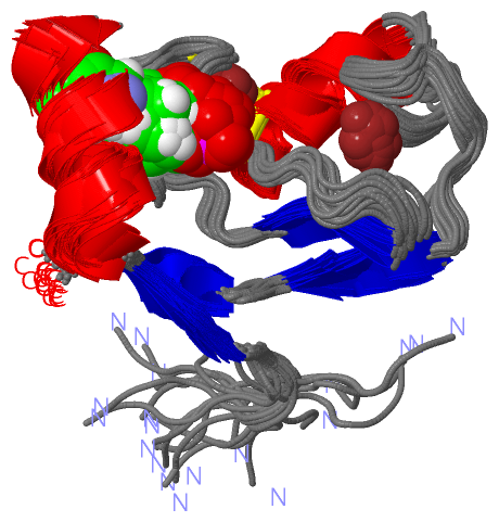
 Visualization

(-) Interactive Views

NMR Structure
  Complete Structure
    Jena3D(integrated viewing of ligand, site, SAP, PROSITE, SCOP information)
    WebMol | AstexViewer[tm]@PDBe
(Java Applets, require no local installation except for Java; loading may be slow)
    STRAP
(Java WebStart application, automatic local installation, requires Java; full application with system access!)
    RasMol
(require local installation)
    Molscript (VRML)
(requires installation of a VRML viewer; select preferred view via VRML and generate a mono or stereo PDF format file)
 
  Ligands, Modified Residues, Ions
    PTR  [ RasMol | Jena3D ]  +environment [ RasMol | Jena3D ]
    ZN  [ RasMol | Jena3D ]  +environment [ RasMol | Jena3D ]
 
  Sites
    AC1  [ RasMol ]  +environment [ RasMol ]
    AC2  [ RasMol ]  +environment [ RasMol ]
 
  Cis Peptide Bonds
    Glu A:386 - Pro A:387   [ RasMol ]  
 

(-) Still Images

Jmol
  protein: cartoon or spacefill or dots and stick; nucleic acid: cartoon and stick; ligands: spacefill; active site: stick
Molscript
  protein, nucleic acid: cartoon; ligands: spacefill; active site: ball and stick

 Databases and Analysis Tools

(-) Databases

Access by PDB/NDB ID
  2ldr
    Family and Domain InformationProDom | SYSTERS
    General Structural InformationGlycoscienceDB | MMDB | NDB | OCA | PDB | PDBe | PDBj | PDBsum | PDBWiki | PQS | PROTEOPEDIA
    Orientation in MembranesOPM
    Protein SurfaceSURFACE
    Secondary StructureDSSP (structure derived) | HSSP (homology derived)
    Structural GenomicsGeneCensus
    Structural NeighboursCE | VAST
    Structure ClassificationCATH | Dali | SCOP
    Validation and Original DataBMRB Data View | BMRB Restraints Grid | EDS | PROCHECK | RECOORD | WHAT_CHECK
 
Access by UniProt ID/Accession number
  CBLB_HUMAN | Q13191
    Comparative Protein Structure ModelsModBase
    Genomic InformationEnsembl
    Protein-protein InteractionDIP
    Sequence, Family and Domain InformationInterPro | Pfam | SMART | UniProtKB/SwissProt
 
Access by Enzyme Classificator   (EC Number)
  6.3.2.-
    General Enzyme InformationBRENDA | EC-PDB | Enzyme | IntEnz
    PathwayKEGG | MetaCyc
 
Access by Disease Identifier   (MIM ID)
  (no 'MIM ID' available)
    Disease InformationOMIM
 
Access by GenAge ID
  (no 'GenAge ID' available)
    Age Related InformationGenAge

(-) Analysis Tools

Access by PDB/NDB ID
    Domain InformationXDom
    Interatomic Contacts of Structural UnitsCSU
    Ligand-protein ContactsLPC
    Protein CavitiescastP
    Sequence and Secondary StructurePDBCartoon
    Structure AlignmentSTRAP(Java WebStart application, automatic local installation, requires Java; full application with system access!)
    Structure and Sequence BrowserSTING
 
Access by UniProt ID/Accession number
  CBLB_HUMAN | Q13191
    Protein Disorder PredictionDisEMBL | FoldIndex | GLOBPLOT (for more information see DisProt)

 Related Entries

(-) Entries Sharing at Least One Protein Chain (UniProt ID)

UniProtKB/Swiss-Prot
        CBLB_HUMAN | Q131912ak5 2bz8 2do6 2j6f 2jnh 2ooa 2oob 3pfv 3vgo 3zni

(-) Related Entries Specified in the PDB File

(no "Related Entries Specified in the PDB File" available for 2LDR)