| biological process |
|---|
| | GO:0015979 | | photosynthesis | | The synthesis by organisms of organic chemical compounds, especially carbohydrates, from carbon dioxide (CO2) using energy obtained from light rather than from the oxidation of chemical compounds. |
| | GO:0010207 | | photosystem II assembly | | The aggregation, arrangement and bonding together of a set of components to form a photosystem II complex on the thylakoid membrane. The photosystem II complex consists of at least 20 polypeptides and around 80 cofactors in most organisms. |
| | GO:0010206 | | photosystem II repair | | Proteolysis of the damaged D1 protein and re-assembly of a new D1 subunit in the photosystem II following photoinhibition. |
| cellular component |
|---|
| | GO:0016020 | | membrane | | A lipid bilayer along with all the proteins and protein complexes embedded in it an attached to it. |
| | GO:0009523 | | photosystem II | | A photosystem that contains a pheophytin-quinone reaction center with associated accessory pigments and electron carriers. In cyanobacteria and chloroplasts, in the presence of light, PSII functions as a water-plastoquinone oxidoreductase, transferring electrons from water to plastoquinone, whereas other photosynthetic bacteria carry out anoxygenic photosynthesis and oxidize other compounds to re-reduce the photoreaction center. |
| | GO:0030096 | | plasma membrane-derived thylakoid photosystem II | | A protein complex, located in the membrane-derived thylakoid, containing the P680 reaction center. In the light, PSII functions as a water-plastoquinone oxidoreductase, transferring electrons from water to plastoquinone. |
| | GO:0009579 | | thylakoid | | A membranous cellular structure that bears the photosynthetic pigments in plants, algae, and cyanobacteria. In cyanobacteria thylakoids are of various shapes and are attached to, or continuous with, the plasma membrane. In eukaryotes they are flattened, membrane-bounded disk-like structures located in the chloroplasts; in the chloroplasts of higher plants the thylakoids form dense stacks called grana. Isolated thylakoid preparations can carry out photosynthetic electron transport and the associated phosphorylation. |
| | GO:0031977 | | thylakoid lumen | | The volume enclosed by a thylakoid membrane. |
| | GO:0042651 | | thylakoid membrane | | The pigmented membrane of any thylakoid. |
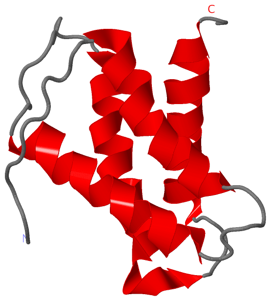
 Description
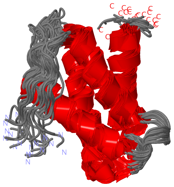