| molecular function |
|---|
| | GO:0017124 | | SH3 domain binding | | Interacting selectively and non-covalently with a SH3 domain (Src homology 3) of a protein, small protein modules containing approximately 50 amino acid residues found in a great variety of intracellular or membrane-associated proteins. |
| | GO:0005515 | | protein binding | | Interacting selectively and non-covalently with any protein or protein complex (a complex of two or more proteins that may include other nonprotein molecules). |
| biological process |
|---|
| | GO:0006915 | | apoptotic process | | A programmed cell death process which begins when a cell receives an internal (e.g. DNA damage) or external signal (e.g. an extracellular death ligand), and proceeds through a series of biochemical events (signaling pathway phase) which trigger an execution phase. The execution phase is the last step of an apoptotic process, and is typically characterized by rounding-up of the cell, retraction of pseudopodes, reduction of cellular volume (pyknosis), chromatin condensation, nuclear fragmentation (karyorrhexis), plasma membrane blebbing and fragmentation of the cell into apoptotic bodies. When the execution phase is completed, the cell has died. |
| | GO:0016477 | | cell migration | | The controlled self-propelled movement of a cell from one site to a destination guided by molecular cues. Cell migration is a central process in the development and maintenance of multicellular organisms. |
| | GO:0007267 | | cell-cell signaling | | Any process that mediates the transfer of information from one cell to another. This process includes signal transduction in the receiving cell and, where applicable, release of a ligand and any processes that actively facilitate its transport and presentation to the receiving cell. Examples include signaling via soluble ligands, via cell adhesion molecules and via gap junctions. |
| | GO:0007010 | | cytoskeleton organization | | A process that is carried out at the cellular level which results in the assembly, arrangement of constituent parts, or disassembly of cytoskeletal structures. |
| | GO:0006897 | | endocytosis | | A vesicle-mediated transport process in which cells take up external materials or membrane constituents by the invagination of a small region of the plasma membrane to form a new membrane-bounded vesicle. |
| | GO:0042059 | | negative regulation of epidermal growth factor receptor signaling pathway | | Any process that stops, prevents, or reduces the frequency, rate or extent of epidermal growth factor receptor signaling pathway activity. |
| | GO:0008360 | | regulation of cell shape | | Any process that modulates the surface configuration of a cell. |
| cellular component |
|---|
| | GO:0030054 | | cell junction | | A cellular component that forms a specialized region of connection between two or more cells or between a cell and the extracellular matrix. At a cell junction, anchoring proteins extend through the plasma membrane to link cytoskeletal proteins in one cell to cytoskeletal proteins in neighboring cells or to proteins in the extracellular matrix. |
| | GO:0005911 | | cell-cell junction | | A cell junction that forms a connection between two or more cells in a multicellular organism; excludes direct cytoplasmic junctions such as ring canals. |
| | GO:0005737 | | cytoplasm | | All of the contents of a cell excluding the plasma membrane and nucleus, but including other subcellular structures. |
| | GO:0031410 | | cytoplasmic vesicle | | A vesicle found in the cytoplasm of a cell. |
| | GO:0030659 | | cytoplasmic vesicle membrane | | The lipid bilayer surrounding a cytoplasmic vesicle. |
| | GO:0005856 | | cytoskeleton | | Any of the various filamentous elements that form the internal framework of cells, and typically remain after treatment of the cells with mild detergent to remove membrane constituents and soluble components of the cytoplasm. The term embraces intermediate filaments, microfilaments, microtubules, the microtrabecular lattice, and other structures characterized by a polymeric filamentous nature and long-range order within the cell. The various elements of the cytoskeleton not only serve in the maintenance of cellular shape but also have roles in other cellular functions, including cellular movement, cell division, endocytosis, and movement of organelles. |
| | GO:0005829 | | cytosol | | The part of the cytoplasm that does not contain organelles but which does contain other particulate matter, such as protein complexes. |
| | GO:0030139 | | endocytic vesicle | | A membrane-bounded intracellular vesicle formed by invagination of the plasma membrane around an extracellular substance. Endocytic vesicles fuse with early endosomes to deliver the cargo for further sorting. |
| | GO:0005925 | | focal adhesion | | Small region on the surface of a cell that anchors the cell to the extracellular matrix and that forms a point of termination of actin filaments. |
| | GO:0016020 | | membrane | | A lipid bilayer along with all the proteins and protein complexes embedded in it an attached to it. |
| | GO:0043005 | | neuron projection | | A prolongation or process extending from a nerve cell, e.g. an axon or dendrite. |
| | GO:0005886 | | plasma membrane | | The membrane surrounding a cell that separates the cell from its external environment. It consists of a phospholipid bilayer and associated proteins. |
| | GO:0045202 | | synapse | | The junction between a nerve fiber of one neuron and another neuron, muscle fiber or glial cell. As the nerve fiber approaches the synapse it enlarges into a specialized structure, the presynaptic nerve ending, which contains mitochondria and synaptic vesicles. At the tip of the nerve ending is the presynaptic membrane; facing it, and separated from it by a minute cleft (the synaptic cleft) is a specialized area of membrane on the receiving cell, known as the postsynaptic membrane. In response to the arrival of nerve impulses, the presynaptic nerve ending secretes molecules of neurotransmitters into the synaptic cleft. These diffuse across the cleft and transmit the signal to the postsynaptic membrane. |
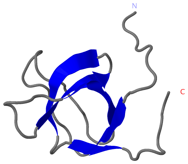
 Description
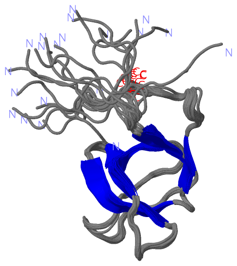