Show PDB file:   
         Plain Text   HTML   (compressed file size)
QuickSearch:   
by PDB,NDB,UniProt,PROSITE Code or Search Term(s)  
(-)NMR Structure - model 1
(-)NMR Structure - all models
collapse expand < >
Image NMR Structure - model 1
NMR Structure - model 1  (Jmol Viewer)
Image NMR Structure - all models
NMR Structure - all models  (Jmol Viewer)

(-) Description

Title :  SOLUTION STRUCTURE OF BB' DOMAINS OF HUMAN PROTEIN DISULFIDE ISOMERASE
 
Authors :  A. Y. Denisov, P. Maattanen, C. Dabrowski, G. Kozlov, D. Y. Thomas, K. Gehring
Date :  22 Feb 08  (Deposition) - 29 Apr 08  (Release) - 10 Mar 09  (Revision)
Method :  SOLUTION NMR
Resolution :  NOT APPLICABLE
Chains :  NMR Structure  :  A  (10x)
Keywords :  Pdi, Endoplasmic Reticulum, Disulfide Bonds, Protein Folding, Chaperone, Isomerase, Membrane, Redox-Active Center (Keyword Search: [Gene Ontology, PubMed, Web (Google))
 
Reference :  A. Y. Denisov, P. Maattanen, C. Dabrowski, G. Kozlov, D. Y. Thomas, K. Gehring
Solution Structure Of The Bb' Domains Of Human Protein Disulfide Isomerase.
Febs J. V. 276 1440 2009
PubMed-ID: 19187238  |  Reference-DOI: 10.1111/J.1742-4658.2009.06884.X
(for further references see the PDB file header)

(-) Compounds

Molecule 1 - PROTEIN DISULFIDE-ISOMERASE
    ChainsA
    EC Number5.3.4.1
    EngineeredYES
    Expression SystemESCHERICHIA COLI BL21
    Expression System PlasmidPGEX-6P1
    Expression System StrainBL21
    Expression System Taxid511693
    Expression System Vector TypePLASMID
    FragmentRESIDUES 135-357
    GeneP4HB, ERBA2L, PDI, PDIA1, PO4DB
    Organism CommonHUMAN
    Organism ScientificHOMO SAPIENS
    Organism Taxid9606
    SynonymPDI, PROLYL 4- HYDROXYLASE SUBUNIT BETA, CELLULAR THYROID HORMONE-BINDING PROTEIN, P55

 Structural Features

(-) Chains, Units

  
NMR Structure (10x)

Summary Information (see also Sequences/Alignments below)

(-) Ligands, Modified Residues, Ions  (0, 0)

(no "Ligand,Modified Residues,Ions" information available for 2K18)

(-) Sites  (0, 0)

(no "Site" information available for 2K18)

(-) SS Bonds  (0, 0)

(no "SS Bond" information available for 2K18)

(-) Cis Peptide Bonds  (0, 0)

(no "Cis Peptide Bond" information available for 2K18)

 Sequence-Structure Mapping

(-) SAPs(SNPs)/Variants  (0, 0)

(no "SAP(SNP)/Variant" information available for 2K18)

(-) PROSITE Motifs  (1, 1)

NMR Structure (1, 1)
 PROSITEUniProtKBPDB
No.IDACDescriptionIDLocationCountLocation
1THIOREDOXIN_2PS51352 Thioredoxin domain profile.PDIA1_HUMAN8-134
349-475
  1-
A:220-228

(-) Exons   (7, 7)

NMR Structure (7, 7)
 ENSEMBLUniProtKBPDB
No.Transcript IDExonExon IDGenome LocationLengthIDLocationLengthCountLocationLength
1.1aENST000003314831aENSE00001316430chr17:79818545-79818203343PDIA1_HUMAN1-49490--
1.3cENST000003314833cENSE00001326286chr17:79817263-79817057207PDIA1_HUMAN49-118701A:1-66
1.4dENST000003314834dENSE00001312577chr17:79813462-79813329134PDIA1_HUMAN118-162451A:7-3327
1.5cENST000003314835cENSE00001754530chr17:79813155-79813018138PDIA1_HUMAN163-208461A:34-7946
1.6bENST000003314836bENSE00001691511chr17:79805223-79805119105PDIA1_HUMAN209-243351A:80-11435
1.7bENST000003314837bENSE00001800920chr17:79804948-79804823126PDIA1_HUMAN244-285421A:115-15642
1.8cENST000003314838cENSE00001661432chr17:79804505-79804305201PDIA1_HUMAN286-352671A:157-22367
1.9aENST000003314839aENSE00001665413chr17:79803867-79803747121PDIA1_HUMAN353-393411A:224-2285
1.9dENST000003314839dENSE00001784888chr17:79803618-79803437182PDIA1_HUMAN393-453610--
1.10cENST0000033148310cENSE00001655750chr17:79803106-7980302087PDIA1_HUMAN454-482290--
1.11dENST0000033148311dENSE00001594571chr17:79801968-79801037932PDIA1_HUMAN483-508260--

(-) Sequences/Alignments

NMR Structure
   Reformat: Number of residues per line =  ('0' or empty: single-line sequence representation)
  Number of residues per labelling interval =   
  UniProt sequence: complete  aligned part    
   Show mapping: SCOP domains CATH domains Pfam domains Secondary structure (by author)
SAPs(SNPs) PROSITE motifs Exons
(details for a mapped element are shown in a popup box when the mouse pointer rests over it)
Chain A from PDB  Type:PROTEIN  Length:228
 aligned with PDIA1_HUMAN | P07237 from UniProtKB/Swiss-Prot  Length:508

    Alignment length:250
                                   117       127       137       147       157       167       177       187       197       207       217       227       237       247       257       267       277       287       297       307       317       327       337       347       357
          PDIA1_HUMAN   108 GDTASPKEYTAGREADDIVNWLKKRTGPAATTLPDGAAAESLVESSEVAVIGFFKDVESDSAKQFLQAAEAIDDIPFGITSNSDVFSKYQLDKDGVVLFKKFDEGRNNFEGEVTKENLLDFIKHNQLPLVIEFTEQTAPKIFGGEIKTHILLFLPKSVSDYDGKLSNFKTAAESFKGKILFIFIDSDHTDNQRILEFFGLKKEECPAVRLITLEEEMTKYKPESEELTAERITEFCHRFLEGKIKPHLMS 357
               SCOP domains ---------------------------------------------------------------------------------------------------------------------------------------------------------------------------------------------------------------------------------------------------------- SCOP domains
               CATH domains ---------------------------------------------------------------------------------------------------------------------------------------------------------------------------------------------------------------------------------------------------------- CATH domains
               Pfam domains -----------------------------------------------------Thioredoxin_6-2k18A01 A:32-216                                                                                                                                                           ------------ Pfam domains
         Sec.struct. author ......----------------------.eee..hhhhhhhhhhh...eeeee.....hhhhhhhhhhhhhh....eeee.hhhhhhhh.....eeeee......eee.....hhhhhhhhhhhhh.....ee..hhhhhhhh....eeeeeee.....hhhhhhhhhhhhhhhh...eeeeeee.....hhhhhhhhh.......eeeeeee...eeee......hhhhhhhhhhhhhhh......... Sec.struct. author
                 SAPs(SNPs) ---------------------------------------------------------------------------------------------------------------------------------------------------------------------------------------------------------------------------------------------------------- SAPs(SNPs)
                    PROSITE THIOREDOXIN_2  PDB: -      ----------------------------------------------------------------------------------------------------------------------------------------------------------------------------------------------------------------------THIOREDOX PROSITE
           Transcript 1 (1) Exon 1.3c  --------------------------------------------Exon 1.5c  PDB: A:34-79 UniProt: 163-208      Exon 1.6b  PDB: A:80-114           Exon 1.7b  PDB: A:115-156 UniProt: 244-285Exon 1.8c  PDB: A:157-223 UniProt: 286-352                         1.9a  Transcript 1 (1)
           Transcript 1 (2) ----------Exon 1.4d  PDB: A:7-33 UniProt: 118-162      --------------------------------------------------------------------------------------------------------------------------------------------------------------------------------------------------- Transcript 1 (2)
                 2k18 A   1 GPLGSP----------------------AATTLPDGAAAESLVESSEVAVIGFFKDVESDSAKQFLQAAEAIDDIPFGITSNSDVFSKYQLDKDGVVLFKKFDEGRNNFEGEVTKENLLDFIKHNQLPLVIEFTEQTAPKIFGGEIKTHILLFLPKSVSDYDGKLSNFKTAAESFKGKILFIFIDSDHTDNQRILEFFGLKKEECPAVRLITLEEEMTKYKPESEELTAERITEFCHRFLEGKIKPHLMS 228
                                 |   -         -        |8        18        28        38        48        58        68        78        88        98       108       118       128       138       148       158       168       178       188       198       208       218       228
                                 6                      7                                                                                                                                                                                                                             

   Legend:   → Mismatch (orange background)
  - → Gap (green background, '-', border residues have a numbering label)
    → Modified Residue (blue background, lower-case, 'x' indicates undefined single-letter code, labelled with number + name)
  x → Chemical Group (purple background, 'x', labelled with number + name, e.g. ACE or NH2)
  extra numbering lines below/above indicate numbering irregularities and modified residue names etc., number ends below/above '|'

 Classification and Annotation

(-) SCOP Domains  (0, 0)

(no "SCOP Domain" information available for 2K18)

(-) CATH Domains  (0, 0)

(no "CATH Domain" information available for 2K18)

(-) Pfam Domains  (1, 1)

NMR Structure

(-) Gene Ontology  (28, 28)

NMR Structure(hide GO term definitions)
Chain A   (PDIA1_HUMAN | P07237)
molecular function
    GO:0019899    enzyme binding    Interacting selectively and non-covalently with any enzyme.
    GO:0005178    integrin binding    Interacting selectively and non-covalently with an integrin.
    GO:0016853    isomerase activity    Catalysis of the geometric or structural changes within one molecule. Isomerase is the systematic name for any enzyme of EC class 5.
    GO:0004656    procollagen-proline 4-dioxygenase activity    Catalysis of the reaction: procollagen L-proline + 2-oxoglutarate + O2 = procollagen trans-4-hydroxy-L-proline + succinate + CO2.
    GO:0005515    protein binding    Interacting selectively and non-covalently with any protein or protein complex (a complex of two or more proteins that may include other nonprotein molecules).
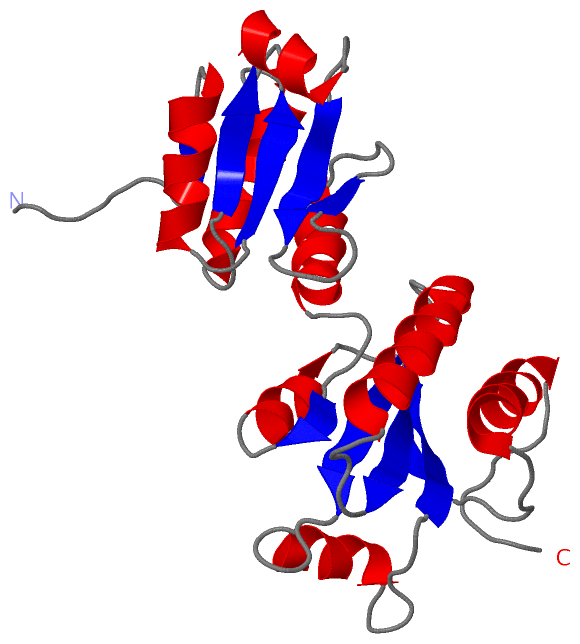
    GO:0003756    protein disulfide isomerase activity    Catalysis of the rearrangement of both intrachain and interchain disulfide bonds in proteins.
    GO:0046982    protein heterodimerization activity    Interacting selectively and non-covalently with a nonidentical protein to form a heterodimer.
biological process
    GO:0045454    cell redox homeostasis    Any process that maintains the redox environment of a cell or compartment within a cell.
    GO:0071456    cellular response to hypoxia    Any process that results in a change in state or activity of a cell (in terms of movement, secretion, enzyme production, gene expression, etc.) as a result of a stimulus indicating lowered oxygen tension. Hypoxia, defined as a decline in O2 levels below normoxic levels of 20.8 - 20.95%, results in metabolic adaptation at both the cellular and organismal level.
    GO:0042158    lipoprotein biosynthetic process    The chemical reactions and pathways resulting in the formation of any conjugated, water-soluble protein in which the covalently attached nonprotein group consists of a lipid or lipids.
    GO:0018401    peptidyl-proline hydroxylation to 4-hydroxy-L-proline    The modification of peptidyl-proline to form 4-hydroxy-L-proline; catalyzed by procollagen-proline,2-oxoglutarate-4-dioxygenase.
    GO:0046598    positive regulation of viral entry into host cell    Any process that activates or increases the frequency, rate or extent of the introduction of viral entry into the host cell.
    GO:0006457    protein folding    The process of assisting in the covalent and noncovalent assembly of single chain polypeptides or multisubunit complexes into the correct tertiary structure.
    GO:1902175    regulation of oxidative stress-induced intrinsic apoptotic signaling pathway    Any process that modulates the frequency, rate or extent of an oxidative stress-induced intrinsic apoptotic signaling pathway.
    GO:0034976    response to endoplasmic reticulum stress    Any process that results in a change in state or activity of a cell (in terms of movement, secretion, enzyme production, gene expression, etc.) as a result of a stress acting at the endoplasmic reticulum. ER stress usually results from the accumulation of unfolded or misfolded proteins in the ER lumen.
    GO:0000302    response to reactive oxygen species    Any process that results in a change in state or activity of a cell or an organism (in terms of movement, secretion, enzyme production, gene expression, etc.) as a result of a reactive oxygen species stimulus. Reactive oxygen species include singlet oxygen, superoxide, and oxygen free radicals.
cellular component
    GO:0005783    endoplasmic reticulum    The irregular network of unit membranes, visible only by electron microscopy, that occurs in the cytoplasm of many eukaryotic cells. The membranes form a complex meshwork of tubular channels, which are often expanded into slitlike cavities called cisternae. The ER takes two forms, rough (or granular), with ribosomes adhering to the outer surface, and smooth (with no ribosomes attached).
    GO:0034663    endoplasmic reticulum chaperone complex    A protein complex that is located in the endoplasmic reticulum and is composed of chaperone proteins, including BiP, GRP94; CaBP1, protein disulfide isomerase (PDI), ERdj3, cyclophilin B, ERp72, GRP170, UDP-glucosyltransferase, and SDF2-L1.
    GO:0005788    endoplasmic reticulum lumen    The volume enclosed by the membranes of the endoplasmic reticulum.
    GO:0005793    endoplasmic reticulum-Golgi intermediate compartment    A complex system of membrane-bounded compartments located between endoplasmic reticulum (ER) and the Golgi complex, with a distinctive membrane protein composition; involved in ER-to-Golgi and Golgi-to-ER transport.
    GO:0009897    external side of plasma membrane    The leaflet of the plasma membrane that faces away from the cytoplasm and any proteins embedded or anchored in it or attached to its surface.
    GO:0070062    extracellular exosome    A vesicle that is released into the extracellular region by fusion of the limiting endosomal membrane of a multivesicular body with the plasma membrane. Extracellular exosomes, also simply called exosomes, have a diameter of about 40-100 nm.
    GO:0005576    extracellular region    The space external to the outermost structure of a cell. For cells without external protective or external encapsulating structures this refers to space outside of the plasma membrane. This term covers the host cell environment outside an intracellular parasite.
    GO:0005925    focal adhesion    Small region on the surface of a cell that anchors the cell to the extracellular matrix and that forms a point of termination of actin filaments.
    GO:0042470    melanosome    A tissue-specific, membrane-bounded cytoplasmic organelle within which melanin pigments are synthesized and stored. Melanosomes are synthesized in melanocyte cells.
    GO:0016020    membrane    A lipid bilayer along with all the proteins and protein complexes embedded in it an attached to it.
    GO:0005886    plasma membrane    The membrane surrounding a cell that separates the cell from its external environment. It consists of a phospholipid bilayer and associated proteins.
    GO:0016222    procollagen-proline 4-dioxygenase complex    A protein complex that catalyzes the formation of procollagen trans-4-hydroxy-L-proline and succinate from procollagen L-proline and 2-oxoglutarate, requiring Fe2+ and ascorbate. Contains two alpha subunits that contribute to most parts of the catalytic sites, and two beta subunits that are identical to protein-disulfide isomerase.

 Visualization

(-) Interactive Views

NMR Structure
  Complete Structure
    Jena3D(integrated viewing of ligand, site, SAP, PROSITE, SCOP information)
    WebMol | AstexViewer[tm]@PDBe
(Java Applets, require no local installation except for Java; loading may be slow)
    STRAP
(Java WebStart application, automatic local installation, requires Java; full application with system access!)
    RasMol
(require local installation)
    Molscript (VRML)
(requires installation of a VRML viewer; select preferred view via VRML and generate a mono or stereo PDF format file)
 
  Ligands, Modified Residues, Ions
(no "Ligands, Modified Residues, Ions" information available for 2k18)
 
  Sites
(no "Sites" information available for 2k18)
 
  Cis Peptide Bonds
(no "Cis Peptide Bonds" information available for 2k18)
 

(-) Still Images

Jmol
  protein: cartoon or spacefill or dots and stick; nucleic acid: cartoon and stick; ligands: spacefill; active site: stick
Molscript
  protein, nucleic acid: cartoon; ligands: spacefill; active site: ball and stick

 Databases and Analysis Tools

(-) Databases

Access by PDB/NDB ID
  2k18
    Family and Domain InformationProDom | SYSTERS
    General Structural InformationGlycoscienceDB | MMDB | NDB | OCA | PDB | PDBe | PDBj | PDBsum | PDBWiki | PQS | PROTEOPEDIA
    Orientation in MembranesOPM
    Protein SurfaceSURFACE
    Secondary StructureDSSP (structure derived) | HSSP (homology derived)
    Structural GenomicsGeneCensus
    Structural NeighboursCE | VAST
    Structure ClassificationCATH | Dali | SCOP
    Validation and Original DataBMRB Data View | BMRB Restraints Grid | EDS | PROCHECK | RECOORD | WHAT_CHECK
 
Access by UniProt ID/Accession number
  PDIA1_HUMAN | P07237
    Comparative Protein Structure ModelsModBase
    Genomic InformationEnsembl
    Protein-protein InteractionDIP
    Sequence, Family and Domain InformationInterPro | Pfam | SMART | UniProtKB/SwissProt
 
Access by Enzyme Classificator   (EC Number)
  5.3.4.1
    General Enzyme InformationBRENDA | EC-PDB | Enzyme | IntEnz
    PathwayKEGG | MetaCyc
 
Access by Disease Identifier   (MIM ID)
  (no 'MIM ID' available)
    Disease InformationOMIM
 
Access by GenAge ID
  (no 'GenAge ID' available)
    Age Related InformationGenAge

(-) Analysis Tools

Access by PDB/NDB ID
    Domain InformationXDom
    Interatomic Contacts of Structural UnitsCSU
    Ligand-protein ContactsLPC
    Protein CavitiescastP
    Sequence and Secondary StructurePDBCartoon
    Structure AlignmentSTRAP(Java WebStart application, automatic local installation, requires Java; full application with system access!)
    Structure and Sequence BrowserSTING
 
Access by UniProt ID/Accession number
  PDIA1_HUMAN | P07237
    Protein Disorder PredictionDisEMBL | FoldIndex | GLOBPLOT (for more information see DisProt)

 Related Entries

(-) Entries Sharing at Least One Protein Chain (UniProt ID)

UniProtKB/Swiss-Prot
        PDIA1_HUMAN | P072371bjx 1mek 1x5c 2bjx 3bj5 3uem 4ekz 4el1 4ju5

(-) Related Entries Specified in the PDB File

1bjx B DOMAIN OF HUMAN PDI