Show PDB file:   
         Plain Text   HTML   (compressed file size)
QuickSearch:   
by PDB,NDB,UniProt,PROSITE Code or Search Term(s)  
(-)Biol.Unit 1 - manually
(-)Asym.Unit - manually
(-)Asymmetric Unit
(-)Asym. Unit - sites
(-)Biological Unit 1
(-)Biol. Unit 1 - sites
collapse expand < >
Image Biol.Unit 1 - manually
Biol.Unit 1 - manually  (Jmol Viewer)
Image Asym.Unit - manually
Asym.Unit - manually  (Jmol Viewer)
Image Asymmetric Unit
Asymmetric Unit  (Jmol Viewer)
Image Asym. Unit - sites
Asym. Unit - sites  (Jmol Viewer)
Image Biological Unit 1
Biological Unit 1  (Jmol Viewer)
Image Biol. Unit 1 - sites
Biol. Unit 1 - sites  (Jmol Viewer)

(-) Description

Title :  CRYSTAL STRUCTURE OF AN XISH FAMILY PROTEIN (ZP_00107633.1) FROM NOSTOC PUNCTIFORME PCC 73102 AT 1.60 A RESOLUTION
 
Authors :  Joint Center For Structural Genomics (Jcsg)
Date :  06 Oct 06  (Deposition) - 24 Oct 06  (Release) - 24 Sep 14  (Revision)
Method :  X-RAY DIFFRACTION
Resolution :  1.60
Chains :  Asym. Unit :  A
Biol. Unit 1:  A  (2x)
Keywords :  Zp_00107633. 1, Hypothetical Protein, Structural Genomics, Psi-2, Protein Structure Initiative, Joint Center For Structural Genomics, Jcsg, Structural Genomics-Unknown Function Complex (Keyword Search: [Gene Ontology, PubMed, Web (Google))
 
Reference :  W. C. Hwang, J. W. Golden, J. Pascual, D. Xu, A. Cheltsov, A. Godzik
Site-Specific Recombination Of Nitrogen-Fixation Genes In Cyanobacteria By Xisf-Xish-Xisi Complex: Structures And Models.
Proteins 2014
PubMed-ID: 25179344  |  Reference-DOI: 10.1002/PROT.24679

(-) Compounds

Molecule 1 - HYPOTHETICAL PROTEIN
    ChainsA
    EngineeredYES
    Expression SystemESCHERICHIA COLI
    Expression System Taxid562
    Expression System Vector TypePLASMID
    GeneZP_00107633.1
    Organism ScientificNOSTOC PUNCTIFORME
    Organism Taxid63737
    StrainPCC 73102

 Structural Features

(-) Chains, Units

  1
Asymmetric Unit A
Biological Unit 1 (2x)A

Summary Information (see also Sequences/Alignments below)

(-) Ligands, Modified Residues, Ions  (3, 5)

Asymmetric Unit (3, 5)
No.NameCountTypeFull Name
1CL1Ligand/IonCHLORIDE ION
2GOL2Ligand/IonGLYCEROL
3MSE2Mod. Amino AcidSELENOMETHIONINE
Biological Unit 1 (2, 8)
No.NameCountTypeFull Name
1CL-1Ligand/IonCHLORIDE ION
2GOL4Ligand/IonGLYCEROL
3MSE4Mod. Amino AcidSELENOMETHIONINE

(-) Sites  (3, 3)

Asymmetric Unit (3, 3)
No.NameEvidenceResiduesDescription
1AC1SOFTWARELEU A:101 , THR A:102 , VAL A:129 , HOH A:167 , HOH A:238BINDING SITE FOR RESIDUE CL A 140
2AC2SOFTWARELYS A:17 , ARG A:87 , VAL A:94 , LEU A:95 , GLN A:131 , VAL A:133 , HOH A:146 , HOH A:168 , HOH A:170 , HOH A:176BINDING SITE FOR RESIDUE GOL A 141
3AC3SOFTWARETRP A:20 , ARG A:50 , GLU A:53 , TRP A:137 , HOH A:234BINDING SITE FOR RESIDUE GOL A 142

(-) SS Bonds  (0, 0)

(no "SS Bond" information available for 2INB)

(-) Cis Peptide Bonds  (1, 1)

Asymmetric Unit
No.Residues
1Asp A:25 -Pro A:26

 Sequence-Structure Mapping

(-) SAPs(SNPs)/Variants  (0, 0)

(no "SAP(SNP)/Variant" information available for 2INB)

(-) PROSITE Motifs  (0, 0)

(no "PROSITE Motif" information available for 2INB)

(-) Exons   (0, 0)

(no "Exon" information available for 2INB)

(-) Sequences/Alignments

Asymmetric Unit
   Reformat: Number of residues per line =  ('0' or empty: single-line sequence representation)
  Number of residues per labelling interval =   
  UniProt sequence: complete  aligned part    
   Show mapping: SCOP domains CATH domains Pfam domains Secondary structure (by author)
SAPs(SNPs) PROSITE motifs Exons
(details for a mapped element are shown in a popup box when the mouse pointer rests over it)
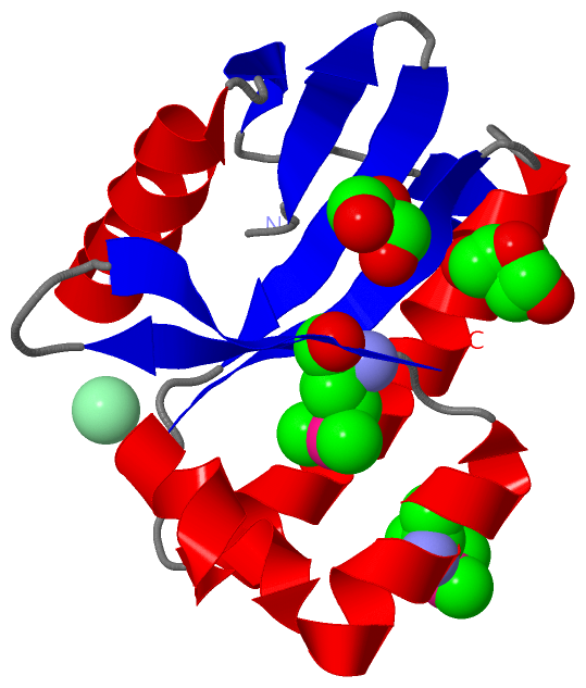
Chain A from PDB  Type:PROTEIN  Length:128
                                                                                                                                                                
               SCOP domains d2inba1 A:5-139 FdxN element excision controlling factor protein                                                                 SCOP domains
               CATH domains 2inbA00 A:5-139  [code=3.40.1350.10, no name defined]                                                                            CATH domains
               Pfam domains -------------------------------------------------------------------------------------------------------------------------------- Pfam domains
         Sec.struct. author hhhhhhhhhhhhhh..eeee.....ee..ee...eeeee..eeeeeee.......hhhhhhhhhhhhhhhhhhhhhh....eeeeeeeehhhhhhhhhhhhhhhhhhhh...eeeee....eeeeee. Sec.struct. author
                 SAPs(SNPs) -------------------------------------------------------------------------------------------------------------------------------- SAPs(SNPs)
                    PROSITE -------------------------------------------------------------------------------------------------------------------------------- PROSITE
                 Transcript -------------------------------------------------------------------------------------------------------------------------------- Transcript
                 2inb A   5 DVFHQVVKIALEKDGWQITNDPLTISVGGVNLKLIAAEREGEKIAVEVKSFLERSSAISEFHTALGQFINYRGALRRRQPERVLYLAVPLTTYKTFFQLDFPKEmIAENQVKmLIYDVEQEVIFQWIN 139
                                    14        24        34 ||     51        61        71        81        91       101       111    |  121  |    131        
                                                          36|                                                                     116-MSE 124-MSE           
                                                           44                                                                                               

   Legend:   → Mismatch (orange background)
  - → Gap (green background, '-', border residues have a numbering label)
    → Modified Residue (blue background, lower-case, 'x' indicates undefined single-letter code, labelled with number + name)
  x → Chemical Group (purple background, 'x', labelled with number + name, e.g. ACE or NH2)
  extra numbering lines below/above indicate numbering irregularities and modified residue names etc., number ends below/above '|'

 Classification and Annotation

(-) SCOP Domains  (1, 1)

Asymmetric Unit

(-) CATH Domains  (1, 1)

Asymmetric Unit
(-)
Class: Alpha Beta (26913)

(-) Pfam Domains  (0, 0)

(no "Pfam Domain" information available for 2INB)

(-) Gene Ontology  (0, 0)

Asymmetric Unit(hide GO term definitions)
    (no "Gene Ontology" information available for 2INB)

 Visualization

(-) Interactive Views

Asymmetric Unit
  Complete Structure
    Jena3D(integrated viewing of ligand, site, SAP, PROSITE, SCOP information)
    WebMol | AstexViewer[tm]@PDBe
(Java Applets, require no local installation except for Java; loading may be slow)
    STRAP
(Java WebStart application, automatic local installation, requires Java; full application with system access!)
    RasMol
(require local installation)
    Molscript (VRML)
(requires installation of a VRML viewer; select preferred view via VRML and generate a mono or stereo PDF format file)
 
  Ligands, Modified Residues, Ions
    CL  [ RasMol | Jena3D ]  +environment [ RasMol | Jena3D ]
    GOL  [ RasMol | Jena3D ]  +environment [ RasMol | Jena3D ]
    MSE  [ RasMol | Jena3D ]  +environment [ RasMol | Jena3D ]
 
  Sites
    AC1  [ RasMol ]  +environment [ RasMol ]
    AC2  [ RasMol ]  +environment [ RasMol ]
    AC3  [ RasMol ]  +environment [ RasMol ]
 
  Cis Peptide Bonds
    Asp A:25 - Pro A:26   [ RasMol ]  
 
Biological Unit
  Complete Structure
    Biological Unit 1  [ Jena3D ]

(-) Still Images

Jmol
  protein: cartoon or spacefill or dots and stick; nucleic acid: cartoon and stick; ligands: spacefill; active site: stick
Molscript
  protein, nucleic acid: cartoon; ligands: spacefill; active site: ball and stick

 Databases and Analysis Tools

(-) Databases

Access by PDB/NDB ID
  2inb
    Family and Domain InformationProDom | SYSTERS
    General Structural InformationGlycoscienceDB | MMDB | NDB | OCA | PDB | PDBe | PDBj | PDBsum | PDBWiki | PQS | PROTEOPEDIA
    Orientation in MembranesOPM
    Protein SurfaceSURFACE
    Secondary StructureDSSP (structure derived) | HSSP (homology derived)
    Structural GenomicsGeneCensus
    Structural NeighboursCE | VAST
    Structure ClassificationCATH | Dali | SCOP
    Validation and Original DataBMRB Data View | BMRB Restraints Grid | EDS | PROCHECK | RECOORD | WHAT_CHECK
 
Access by UniProt ID/Accession number
  (no 'UniProt ID/Accession number' available) |
    Comparative Protein Structure ModelsModBase
    Genomic InformationEnsembl
    Protein-protein InteractionDIP
    Sequence, Family and Domain InformationInterPro | Pfam | SMART | UniProt
 
Access by Enzyme Classificator   (EC Number)
  (no 'Enzyme Classificator' available)
    General Enzyme InformationBRENDA | EC-PDB | Enzyme | IntEnz
    PathwayKEGG | MetaCyc
 
Access by Disease Identifier   (MIM ID)
  (no 'MIM ID' available)
    Disease InformationOMIM
 
Access by GenAge ID
  (no 'GenAge ID' available)
    Age Related InformationGenAge

(-) Analysis Tools

Access by PDB/NDB ID
    Domain InformationXDom
    Interatomic Contacts of Structural UnitsCSU
    Ligand-protein ContactsLPC
    Protein CavitiescastP
    Sequence and Secondary StructurePDBCartoon
    Structure AlignmentSTRAP(Java WebStart application, automatic local installation, requires Java; full application with system access!)
    Structure and Sequence BrowserSTING
 
Access by UniProt ID/Accession number
  (no 'UniProt ID/Accession number' available) |
    Protein Disorder PredictionDisEMBL | FoldIndex | GLOBPLOT

 Related Entries

(-) Entries Sharing at Least One Protein Chain (UniProt ID)

(no "Entries Sharing at Least One Protein Chain" available for 2INB)

(-) Related Entries Specified in the PDB File

(no "Related Entries Specified in the PDB File" available for 2INB)