| molecular function |
|---|
| | GO:0005518 | | collagen binding | | Interacting selectively and non-covalently with collagen, a group of fibrous proteins of very high tensile strength that form the main component of connective tissue in animals. Collagen is highly enriched in glycine (some regions are 33% glycine) and proline, occurring predominantly as 3-hydroxyproline (about 20%). |
| | GO:0004896 | | cytokine receptor activity | | Combining with a cytokine and transmitting the signal from one side of the membrane to the other to initiate a change in cell activity. |
| | GO:0005540 | | hyaluronic acid binding | | Interacting selectively and non-covalently with hyaluronic acid, a polymer composed of repeating dimeric units of glucuronic acid and N-acetyl glucosamine. |
| | GO:0004415 | | hyalurononglucosaminidase activity | | Catalysis of the random hydrolysis of (1->4) linkages between N-acetyl-beta-D-glucosamine and D-glucuronate residues in hyaluronate. |
| | GO:0005515 | | protein binding | | Interacting selectively and non-covalently with any protein or protein complex (a complex of two or more proteins that may include other nonprotein molecules). |
| biological process |
|---|
| | GO:0051216 | | cartilage development | | The process whose specific outcome is the progression of a cartilage element over time, from its formation to the mature structure. Cartilage elements are skeletal elements that consist of connective tissue dominated by extracellular matrix containing collagen type II and large amounts of proteoglycan, particularly chondroitin sulfate. |
| | GO:0007155 | | cell adhesion | | The attachment of a cell, either to another cell or to an underlying substrate such as the extracellular matrix, via cell adhesion molecules. |
| | GO:0007160 | | cell-matrix adhesion | | The binding of a cell to the extracellular matrix via adhesion molecules. |
| | GO:0044344 | | cellular response to fibroblast growth factor stimulus | | Any process that results in a change in state or activity of a cell (in terms of movement, secretion, enzyme production, gene expression, etc.) as a result of an fibroblast growth factor stimulus. |
| | GO:0022617 | | extracellular matrix disassembly | | A process that results in the breakdown of the extracellular matrix. |
| | GO:0030198 | | extracellular matrix organization | | A process that is carried out at the cellular level which results in the assembly, arrangement of constituent parts, or disassembly of an extracellular matrix. |
| | GO:0030214 | | hyaluronan catabolic process | | The chemical reactions and pathways resulting in the breakdown of hyaluronan, the naturally occurring anionic form of hyaluronic acid, any member of a group of glycosaminoglycans, the repeat units of which consist of beta-1,4 linked D-glucuronyl-beta-(1,3)-N-acetyl-D-glucosamine. |
| | GO:0060333 | | interferon-gamma-mediated signaling pathway | | A series of molecular signals initiated by the binding of interferon-gamma to a receptor on the surface of a cell, and ending with regulation of a downstream cellular process, e.g. transcription. Interferon gamma is the only member of the type II interferon found so far. |
| | GO:0050900 | | leukocyte migration | | The movement of a leukocyte within or between different tissues and organs of the body. |
| | GO:0070487 | | monocyte aggregation | | The adhesion of one monocyte to one or more other monocytes via adhesion molecules. |
| | GO:0043518 | | negative regulation of DNA damage response, signal transduction by p53 class mediator | | Any process that stops, prevents, or reduces the frequency, rate or extent of the cascade of processes induced by the cell cycle regulator phosphoprotein p53, or an equivalent protein, in response to the detection of DNA damage. |
| | GO:0043066 | | negative regulation of apoptotic process | | Any process that stops, prevents, or reduces the frequency, rate or extent of cell death by apoptotic process. |
| | GO:0043154 | | negative regulation of cysteine-type endopeptidase activity involved in apoptotic process | | Any process that stops, prevents, or reduces the frequency, rate or extent of a cysteine-type endopeptidase activity involved in the apoptotic process. |
| | GO:1902166 | | negative regulation of intrinsic apoptotic signaling pathway in response to DNA damage by p53 class mediator | | Any process that stops, prevents or reduces the frequency, rate or extent of intrinsic apoptotic signaling pathway in response to DNA damage by p53 class mediator. |
| | GO:0070374 | | positive regulation of ERK1 and ERK2 cascade | | Any process that activates or increases the frequency, rate or extent of signal transduction mediated by the ERK1 and ERK2 cascade. |
| | GO:0034116 | | positive regulation of heterotypic cell-cell adhesion | | Any process that activates or increases the frequency, rate, or extent of heterotypic cell-cell adhesion. |
| | GO:1900625 | | positive regulation of monocyte aggregation | | Any process that activates or increases the frequency, rate or extent of monocyte aggregation. |
| | GO:0033138 | | positive regulation of peptidyl-serine phosphorylation | | Any process that activates or increases the frequency, rate or extent of the phosphorylation of peptidyl-serine. |
| | GO:0050731 | | positive regulation of peptidyl-tyrosine phosphorylation | | Any process that activates or increases the frequency, rate or extent of the phosphorylation of peptidyl-tyrosine. |
| | GO:0016337 | | single organismal cell-cell adhesion | | The attachment of one cell to another cell via adhesion molecules, where both cells are part of the same organism. |
| cellular component |
|---|
| | GO:0005794 | | Golgi apparatus | | A compound membranous cytoplasmic organelle of eukaryotic cells, consisting of flattened, ribosome-free vesicles arranged in a more or less regular stack. The Golgi apparatus differs from the endoplasmic reticulum in often having slightly thicker membranes, appearing in sections as a characteristic shallow semicircle so that the convex side (cis or entry face) abuts the endoplasmic reticulum, secretory vesicles emerging from the concave side (trans or exit face). In vertebrate cells there is usually one such organelle, while in invertebrates and plants, where they are known usually as dictyosomes, there may be several scattered in the cytoplasm. The Golgi apparatus processes proteins produced on the ribosomes of the rough endoplasmic reticulum; such processing includes modification of the core oligosaccharides of glycoproteins, and the sorting and packaging of proteins for transport to a variety of cellular locations. Three different regions of the Golgi are now recognized both in terms of structure and function: cis, in the vicinity of the cis face, trans, in the vicinity of the trans face, and medial, lying between the cis and trans regions. |
| | GO:0009986 | | cell surface | | The external part of the cell wall and/or plasma membrane. |
| | GO:0005737 | | cytoplasm | | All of the contents of a cell excluding the plasma membrane and nucleus, but including other subcellular structures. |
| | GO:0070062 | | extracellular exosome | | A vesicle that is released into the extracellular region by fusion of the limiting endosomal membrane of a multivesicular body with the plasma membrane. Extracellular exosomes, also simply called exosomes, have a diameter of about 40-100 nm. |
| | GO:0005925 | | focal adhesion | | Small region on the surface of a cell that anchors the cell to the extracellular matrix and that forms a point of termination of actin filaments. |
| | GO:0016021 | | integral component of membrane | | The component of a membrane consisting of the gene products and protein complexes having at least some part of their peptide sequence embedded in the hydrophobic region of the membrane. |
| | GO:0005887 | | integral component of plasma membrane | | The component of the plasma membrane consisting of the gene products and protein complexes having at least some part of their peptide sequence embedded in the hydrophobic region of the membrane. |
| | GO:0035692 | | macrophage migration inhibitory factor receptor complex | | A protein complex that binds macrophage migration inhibitory factor. Comprises CD74 and CD44 cell surface proteins. |
| | GO:0016020 | | membrane | | A lipid bilayer along with all the proteins and protein complexes embedded in it an attached to it. |
| | GO:0005886 | | plasma membrane | | The membrane surrounding a cell that separates the cell from its external environment. It consists of a phospholipid bilayer and associated proteins. |
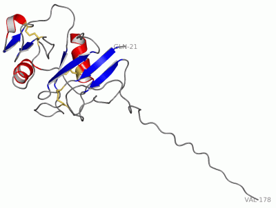
 Description
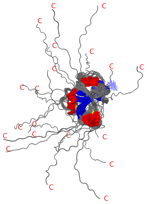