Show PDB file:   
         Plain Text   HTML   (compressed file size)
QuickSearch:   
by PDB,NDB,UniProt,PROSITE Code or Search Term(s)  
(-)Asymmetric Unit
(-)Asym. Unit - sites
(-)Biological Unit 1
(-)Biol. Unit 1 - sites
(-)Biological Unit 2
collapse expand < >
Image Asymmetric Unit
Asymmetric Unit  (Jmol Viewer)
Image Asym. Unit - sites
Asym. Unit - sites  (Jmol Viewer)
Image Biological Unit 1
Biological Unit 1  (Jmol Viewer)
Image Biol. Unit 1 - sites
Biol. Unit 1 - sites  (Jmol Viewer)
Image Biological Unit 2
Biological Unit 2  (Jmol Viewer)

(-) Description

Title :  CRYSTAL STRUCTURES OF THE INTERACTING DOMAINS FROM YEAST GLUTAMYL-TRNA SYNTHETASE AND TRNA AMINOACYLATION AND NUCLEAR EXPORT COFACTOR ARC1P REVEAL A NOVEL FUNCTION FOR AN OLD FOLD
 
Authors :  H. Simader, M. Hothorn, D. Suck
Date :  20 Jul 06  (Deposition) - 05 Sep 06  (Release) - 13 Jul 11  (Revision)
Method :  X-RAY DIFFRACTION
Resolution :  1.90
Chains :  Asym. Unit :  A,B
Biol. Unit 1:  A  (1x)
Biol. Unit 2:  B  (1x)
Keywords :  Gst-Fold, Ligase (Keyword Search: [Gene Ontology, PubMed, Web (Google))
 
Reference :  H. Simader, M. Hothorn, D. Suck
Structures Of The Interacting Domains From Yeast Glutamyl-Trna Synthetase And Trna-Aminoacylation And Nuclear-Export Cofactor Arc1P Reveal A Novel Function For A Old Fold.
Acta Crystallogr. , Sect. D V. 62 1510 2006
PubMed-ID: 17139087  |  Reference-DOI: 10.1107/S0907444906039850
(for further references see the PDB file header)

(-) Compounds

Molecule 1 - GLUTAMYL-TRNA SYNTHETASE, CYTOPLASMIC
    ChainsA, B
    EC Number6.1.1.17
    EngineeredYES
    Expression SystemESCHERICHIA COLI
    Expression System StrainBL21 DE3 STAR
    Expression System Taxid562
    Expression System VectorPETM-DERIVATIVE
    Expression System Vector TypePLASMID
    FragmentN-TERMINAL HETEROMERISATION DOMAIN, RESIDUES 1-207
    GeneGUS1
    Organism CommonBAKER'S YEAST
    Organism ScientificSACCHAROMYCES CEREVISIAE
    Organism Taxid4932
    SynonymGLUTAMATE-TRNA LIGASE, GLURS, P85

 Structural Features

(-) Chains, Units

  12
Asymmetric Unit AB
Biological Unit 1 (1x)A 
Biological Unit 2 (1x) B

Summary Information (see also Sequences/Alignments below)

(-) Ligands, Modified Residues, Ions  (1, 44)

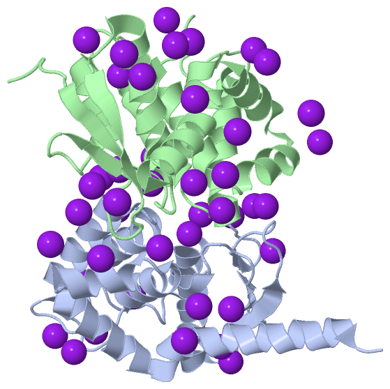
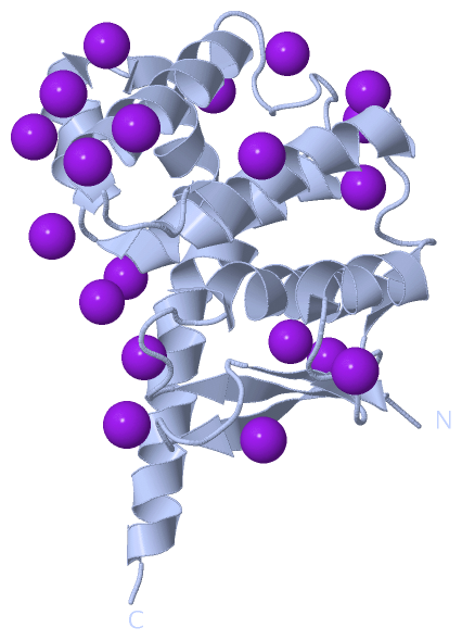
Asymmetric Unit (1, 44)
No.NameCountTypeFull Name
1IOD44Ligand/IonIODIDE ION
Biological Unit 1 (1, 20)
No.NameCountTypeFull Name
1IOD20Ligand/IonIODIDE ION
Biological Unit 2 (1, 24)
No.NameCountTypeFull Name
1IOD24Ligand/IonIODIDE ION

(-) Sites  (30, 30)

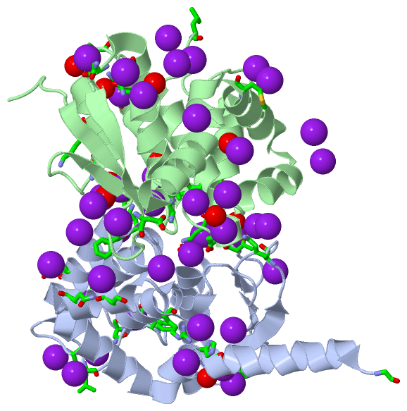
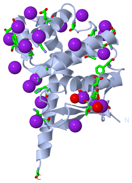
Asymmetric Unit (30, 30)
No.NameEvidenceResiduesDescription
01AC1SOFTWAREPHE A:109BINDING SITE FOR RESIDUE IOD A 402
02AC2SOFTWAREPHE A:85 , ASN A:87 , ALA A:137BINDING SITE FOR RESIDUE IOD A 406
03AC3SOFTWAREPHE B:85 , ALA B:137 , IOD B:421BINDING SITE FOR RESIDUE IOD B 407
04AC4SOFTWAREGLU A:117 , LYS B:76BINDING SITE FOR RESIDUE IOD A 408
05AC5SOFTWARELEU B:133BINDING SITE FOR RESIDUE IOD B 410
06AC6SOFTWARESER A:154 , ASN A:158BINDING SITE FOR RESIDUE IOD A 412
07AC7SOFTWARELYS A:59BINDING SITE FOR RESIDUE IOD A 413
08AC8SOFTWAREASN B:108 , HOH B:478BINDING SITE FOR RESIDUE IOD B 414
09AC9SOFTWAREASN A:108 , ILE B:31BINDING SITE FOR RESIDUE IOD B 415
10BC1SOFTWARETHR A:70 , GLU A:71 , HOH A:554BINDING SITE FOR RESIDUE IOD A 416
11BC2SOFTWAREASN B:49BINDING SITE FOR RESIDUE IOD B 417
12BC3SOFTWAREASN A:44 , GLY A:198 , HOH A:483 , HOH A:563BINDING SITE FOR RESIDUE IOD A 418
13BC4SOFTWAREASP A:72 , LYS B:111 , GLN B:114BINDING SITE FOR RESIDUE IOD B 419
14BC5SOFTWAREIOD B:423BINDING SITE FOR RESIDUE IOD B 420
15BC6SOFTWAREPHE B:85 , LYS B:90 , IOD B:407 , HOH B:541BINDING SITE FOR RESIDUE IOD B 421
16BC7SOFTWAREVAL B:32 , SER B:148BINDING SITE FOR RESIDUE IOD B 422
17BC8SOFTWAREIOD B:420BINDING SITE FOR RESIDUE IOD B 423
18BC9SOFTWARELYS A:111 , ASP B:72BINDING SITE FOR RESIDUE IOD A 424
19CC1SOFTWAREASN B:44 , HOH B:524BINDING SITE FOR RESIDUE IOD B 426
20CC2SOFTWAREASN A:49BINDING SITE FOR RESIDUE IOD A 428
21CC3SOFTWAREARG A:147 , HIS A:181BINDING SITE FOR RESIDUE IOD A 430
22CC4SOFTWARETHR A:127BINDING SITE FOR RESIDUE IOD A 431
23CC5SOFTWAREGLU B:103 , HOH B:477BINDING SITE FOR RESIDUE IOD B 432
24CC6SOFTWAREASN A:108 , HOH B:476 , HOH B:495BINDING SITE FOR RESIDUE IOD A 433
25CC7SOFTWAREASP A:89BINDING SITE FOR RESIDUE IOD A 436
26CC8SOFTWAREASP B:57BINDING SITE FOR RESIDUE IOD B 438
27CC9SOFTWAREHOH B:451BINDING SITE FOR RESIDUE IOD B 439
28DC1SOFTWARELEU A:133 , ALA B:83 , HOH B:518BINDING SITE FOR RESIDUE IOD B 440
29DC2SOFTWAREMET B:173BINDING SITE FOR RESIDUE IOD B 441
30DC3SOFTWAREHOH B:510BINDING SITE FOR RESIDUE IOD B 444

(-) SS Bonds  (0, 0)

(no "SS Bond" information available for 2HRA)

(-) Cis Peptide Bonds  (0, 0)

(no "Cis Peptide Bond" information available for 2HRA)

 Sequence-Structure Mapping

(-) SAPs(SNPs)/Variants  (0, 0)

(no "SAP(SNP)/Variant" information available for 2HRA)

(-) PROSITE Motifs  (0, 0)

(no "PROSITE Motif" information available for 2HRA)

(-) Exons   (1, 2)

Asymmetric Unit (1, 2)
 ENSEMBLUniProtKBPDB
No.Transcript IDExonExon IDGenome LocationLengthIDLocationLengthCountLocationLength
1.1YGL245W1YGL245W.1VII:39023-411492127SYEC_YEAST1-7087082A:19-198
B:19-193
180
175

(-) Sequences/Alignments

Asymmetric Unit
   Reformat: Number of residues per line =  ('0' or empty: single-line sequence representation)
  Number of residues per labelling interval =   
  UniProt sequence: complete  aligned part    
   Show mapping: SCOP domains CATH domains Pfam domains Secondary structure (by author)
SAPs(SNPs) PROSITE motifs Exons
(details for a mapped element are shown in a popup box when the mouse pointer rests over it)
Chain A from PDB  Type:PROTEIN  Length:180
 aligned with SYEC_YEAST | P46655 from UniProtKB/Swiss-Prot  Length:708

    Alignment length:180
                                    10        20        30        40        50        60        70        80        90       100       110       120       130       140       150       160       170       180
           SYEC_YEAST     1 MPSTLTINGKAPIVAYAELIAARIVNALAPNSIAIKLVDDKKAPAAKLDDATEDVFNKITSKFAAIFDNGDKEQVAKWVNLAQKELVIKNFAKLSQSLETLDSQLNLRTFILGGLKYSAADVACWGALRSNGMCGSIIKNKVDVNVSRWYTLLEMDPIFGEAHDFLSKSLLELKKSANVG 180
               SCOP domains ------------------------------------------------------------------------------------------------------------------------------------------------------------------------------------ SCOP domains
               CATH domains ------------------------------------------------------------------------------------------------------------------------------------------------------------------------------------ CATH domains
               Pfam domains ------------------------------------------------------------------------------------------------------------------------------------------------------------------------------------ Pfam domains
         Sec.struct. author ..eeeeee......hhhhhhhhhhhhhhh...eeeeee.......eee.....hhhhhhhhhh.......hhhhhhhhhhhhhhh....hhhhhhhhhhhhhhhh............hhhhhhhhhhhhhh.hhhhhhhhh.hhhhhhhhhhhhhhhhhhhhhhhhhhhhhhhhhhhh.. Sec.struct. author
                 SAPs(SNPs) ------------------------------------------------------------------------------------------------------------------------------------------------------------------------------------ SAPs(SNPs)
                    PROSITE ------------------------------------------------------------------------------------------------------------------------------------------------------------------------------------ PROSITE
               Transcript 1 Exon 1.1  PDB: A:19-198 UniProt: 1-708 [INCOMPLETE]                                                                                                                                  Transcript 1
                 2hra A  19 MPSTLTINGKAPIVAYAELIAARIVNALAPNSIAIKLVDDKKAPAAKLDDATEDVFNKITSKFAAIFDNGDKEQVAKWVNLAQKELVIKNFAKLSQSLETLDSQLNLRTFILGGLKYSAADVACWGALRSNGMCGSIIKNKVDVNVSRWYTLLEMDPIFGEAHDFLSKSLLELKKSANVG 198
                                    28        38        48        58        68        78        88        98       108       118       128       138       148       158       168       178       188       198

Chain B from PDB  Type:PROTEIN  Length:178
 aligned with SYEC_YEAST | P46655 from UniProtKB/Swiss-Prot  Length:708

    Alignment length:178
                               1                                                                                                                                                                              
                               |     7        17        27        37        47        57        67        77        87        97       107       117       127       137       147       157       167        
           SYEC_YEAST     - ---MPSTLTINGKAPIVAYAELIAARIVNALAPNSIAIKLVDDKKAPAAKLDDATEDVFNKITSKFAAIFDNGDKEQVAKWVNLAQKELVIKNFAKLSQSLETLDSQLNLRTFILGGLKYSAADVACWGALRSNGMCGSIIKNKVDVNVSRWYTLLEMDPIFGEAHDFLSKSLLELKK 175
               SCOP domains ---------------------------------------------------------------------------------------------------------------------------------------------------------------------------------- SCOP domains
               CATH domains ---------------------------------------------------------------------------------------------------------------------------------------------------------------------------------- CATH domains
               Pfam domains ---------------------------------------------------------------------------------------------------------------------------------------------------------------------------------- Pfam domains
         Sec.struct. author .....eeeeee......hhhhhhhhhhhhhhh...eeeeee.......eee.....hhhhhhhhhh.......hhhhhhhhhhhhhhh....hhhhhhhhhhhhhhhh............hhhhhhhhhhhhhh.hhhhhhhhhhhhhhhhhhhhhhhhhhhhhhhhhhhhhhhhhh. Sec.struct. author
                 SAPs(SNPs) ---------------------------------------------------------------------------------------------------------------------------------------------------------------------------------- SAPs(SNPs)
                    PROSITE ---------------------------------------------------------------------------------------------------------------------------------------------------------------------------------- PROSITE
               Transcript 1 ---Exon 1.1  PDB: B:19-193 UniProt: 1-708 [INCOMPLETE]                                                                                                                             Transcript 1
                 2hra B  16 GIKMPSTLTINGKAPIVAYAELIAARIVNALAPNSIAIKLVDDKKAPAAKLDDATEDVFNKITSKFAAIFDNGDKEQVAKWVNLAQKELVIKNFAKLSQSLETLDSQLNLRTFILGGLKYSAADVACWGALRSNGMCGSIIKNKVDVNVSRWYTLLEMDPIFGEAHDFLSKSLLELKK 193
                                    25        35        45        55        65        75        85        95       105       115       125       135       145       155       165       175       185        

   Legend:   → Mismatch (orange background)
  - → Gap (green background, '-', border residues have a numbering label)
    → Modified Residue (blue background, lower-case, 'x' indicates undefined single-letter code, labelled with number + name)
  x → Chemical Group (purple background, 'x', labelled with number + name, e.g. ACE or NH2)
  extra numbering lines below/above indicate numbering irregularities and modified residue names etc., number ends below/above '|'

 Classification and Annotation

(-) SCOP Domains  (0, 0)

(no "SCOP Domain" information available for 2HRA)

(-) CATH Domains  (0, 0)

(no "CATH Domain" information available for 2HRA)

(-) Pfam Domains  (0, 0)

(no "Pfam Domain" information available for 2HRA)

(-) Gene Ontology  (17, 17)

Asymmetric Unit(hide GO term definitions)
Chain A,B   (SYEC_YEAST | P46655)
molecular function
    GO:0005524    ATP binding    Interacting selectively and non-covalently with ATP, adenosine 5'-triphosphate, a universally important coenzyme and enzyme regulator.
    GO:0004812    aminoacyl-tRNA ligase activity    Catalysis of the formation of aminoacyl-tRNA from ATP, amino acid, and tRNA with the release of diphosphate and AMP.
    GO:0004818    glutamate-tRNA ligase activity    Catalysis of the reaction: ATP + L-glutamate + tRNA(Glu) = AMP + diphosphate + L-glutamyl-tRNA(Glu).
    GO:0004819    glutamine-tRNA ligase activity    Catalysis of the reaction: ATP + L-glutamine + tRNA(Gln) = AMP + diphosphate + L-glutaminyl-tRNA(Gln).
    GO:0016874    ligase activity    Catalysis of the joining of two substances, or two groups within a single molecule, with the concomitant hydrolysis of the diphosphate bond in ATP or a similar triphosphate.
    GO:0016876    ligase activity, forming aminoacyl-tRNA and related compounds    Catalysis of the joining of an amino acid and a nucleic acid (usually tRNA) or poly(ribitol phosphate), with the concomitant hydrolysis of the diphosphate bond in ATP or a similar triphosphate. The reaction forms an aminoacyl-tRNA or a related compound.
    GO:0000166    nucleotide binding    Interacting selectively and non-covalently with a nucleotide, any compound consisting of a nucleoside that is esterified with (ortho)phosphate or an oligophosphate at any hydroxyl group on the ribose or deoxyribose.
    GO:0005515    protein binding    Interacting selectively and non-covalently with any protein or protein complex (a complex of two or more proteins that may include other nonprotein molecules).
biological process
    GO:0006425    glutaminyl-tRNA aminoacylation    The process of coupling glutamine to glutaminyl-tRNA, catalyzed by glutaminyl-tRNA synthetase. In tRNA aminoacylation, the amino acid is first activated by linkage to AMP and then transferred to either the 2'- or the 3'-hydroxyl group of the 3'- adenosine residue of the tRNA.
    GO:0006424    glutamyl-tRNA aminoacylation    The process of coupling glutamate to glutamyl-tRNA, catalyzed by glutamyl-tRNA synthetase. In tRNA aminoacylation, the amino acid is first activated by linkage to AMP and then transferred to either the 2'- or the 3'-hydroxyl group of the 3'- adenosine residue of the tRNA.
    GO:0043039    tRNA aminoacylation    The chemical reactions and pathways by which the various amino acids become bonded to their corresponding tRNAs. The most common route for synthesis of aminoacyl tRNA is by the formation of an ester bond between the 3'-hydroxyl group of the most 3' adenosine of the tRNA, usually catalyzed by the cognate aminoacyl-tRNA ligase. A given aminoacyl-tRNA ligase aminoacylates all species of an isoaccepting group of tRNA molecules.
    GO:0006418    tRNA aminoacylation for protein translation    The synthesis of aminoacyl tRNA by the formation of an ester bond between the 3'-hydroxyl group of the most 3' adenosine of the tRNA, to be used in ribosome-mediated polypeptide synthesis.
    GO:0006412    translation    The cellular metabolic process in which a protein is formed, using the sequence of a mature mRNA or circRNA molecule to specify the sequence of amino acids in a polypeptide chain. Translation is mediated by the ribosome, and begins with the formation of a ternary complex between aminoacylated initiator methionine tRNA, GTP, and initiation factor 2, which subsequently associates with the small subunit of the ribosome and an mRNA or circRNA. Translation ends with the release of a polypeptide chain from the ribosome.
cellular component
    GO:0005737    cytoplasm    All of the contents of a cell excluding the plasma membrane and nucleus, but including other subcellular structures.
    GO:0005829    cytosol    The part of the cytoplasm that does not contain organelles but which does contain other particulate matter, such as protein complexes.
    GO:0017102    methionyl glutamyl tRNA synthetase complex    A complex consisting of methionyl- and glutamyl-tRNA synthetases. The tRNA synthetases present in the complex bind to their cognate tRNAs more efficiently than they do as monomers.
    GO:0005739    mitochondrion    A semiautonomous, self replicating organelle that occurs in varying numbers, shapes, and sizes in the cytoplasm of virtually all eukaryotic cells. It is notably the site of tissue respiration.

 Visualization

(-) Interactive Views

Asymmetric Unit
  Complete Structure
    Jena3D(integrated viewing of ligand, site, SAP, PROSITE, SCOP information)
    WebMol | AstexViewer[tm]@PDBe
(Java Applets, require no local installation except for Java; loading may be slow)
    STRAP
(Java WebStart application, automatic local installation, requires Java; full application with system access!)
    RasMol
(require local installation)
    Molscript (VRML)
(requires installation of a VRML viewer; select preferred view via VRML and generate a mono or stereo PDF format file)
 
  Ligands, Modified Residues, Ions
    IOD  [ RasMol | Jena3D ]  +environment [ RasMol | Jena3D ]
 
  Sites
    AC1  [ RasMol ]  +environment [ RasMol ]
    AC2  [ RasMol ]  +environment [ RasMol ]
    AC3  [ RasMol ]  +environment [ RasMol ]
    AC4  [ RasMol ]  +environment [ RasMol ]
    AC5  [ RasMol ]  +environment [ RasMol ]
    AC6  [ RasMol ]  +environment [ RasMol ]
    AC7  [ RasMol ]  +environment [ RasMol ]
    AC8  [ RasMol ]  +environment [ RasMol ]
    AC9  [ RasMol ]  +environment [ RasMol ]
    BC1  [ RasMol ]  +environment [ RasMol ]
    BC2  [ RasMol ]  +environment [ RasMol ]
    BC3  [ RasMol ]  +environment [ RasMol ]
    BC4  [ RasMol ]  +environment [ RasMol ]
    BC5  [ RasMol ]  +environment [ RasMol ]
    BC6  [ RasMol ]  +environment [ RasMol ]
    BC7  [ RasMol ]  +environment [ RasMol ]
    BC8  [ RasMol ]  +environment [ RasMol ]
    BC9  [ RasMol ]  +environment [ RasMol ]
    CC1  [ RasMol ]  +environment [ RasMol ]
    CC2  [ RasMol ]  +environment [ RasMol ]
    CC3  [ RasMol ]  +environment [ RasMol ]
    CC4  [ RasMol ]  +environment [ RasMol ]
    CC5  [ RasMol ]  +environment [ RasMol ]
    CC6  [ RasMol ]  +environment [ RasMol ]
    CC7  [ RasMol ]  +environment [ RasMol ]
    CC8  [ RasMol ]  +environment [ RasMol ]
    CC9  [ RasMol ]  +environment [ RasMol ]
    DC1  [ RasMol ]  +environment [ RasMol ]
    DC2  [ RasMol ]  +environment [ RasMol ]
    DC3  [ RasMol ]  +environment [ RasMol ]
 
  Cis Peptide Bonds
(no "Cis Peptide Bonds" information available for 2hra)
 
Biological Units
  Complete Structure
    Biological Unit 1  [ Jena3D ]
    Biological Unit 2  [ Jena3D ]

(-) Still Images

Jmol
  protein: cartoon or spacefill or dots and stick; nucleic acid: cartoon and stick; ligands: spacefill; active site: stick
Molscript
  protein, nucleic acid: cartoon; ligands: spacefill; active site: ball and stick

 Databases and Analysis Tools

(-) Databases

Access by PDB/NDB ID
  2hra
    Family and Domain InformationProDom | SYSTERS
    General Structural InformationGlycoscienceDB | MMDB | NDB | OCA | PDB | PDBe | PDBj | PDBsum | PDBWiki | PQS | PROTEOPEDIA
    Orientation in MembranesOPM
    Protein SurfaceSURFACE
    Secondary StructureDSSP (structure derived) | HSSP (homology derived)
    Structural GenomicsGeneCensus
    Structural NeighboursCE | VAST
    Structure ClassificationCATH | Dali | SCOP
    Validation and Original DataBMRB Data View | BMRB Restraints Grid | EDS | PROCHECK | RECOORD | WHAT_CHECK
 
Access by UniProt ID/Accession number
  SYEC_YEAST | P46655
    Comparative Protein Structure ModelsModBase
    Genomic InformationEnsembl
    Protein-protein InteractionDIP
    Sequence, Family and Domain InformationInterPro | Pfam | SMART | UniProtKB/SwissProt
 
Access by Enzyme Classificator   (EC Number)
  6.1.1.17
    General Enzyme InformationBRENDA | EC-PDB | Enzyme | IntEnz
    PathwayKEGG | MetaCyc
 
Access by Disease Identifier   (MIM ID)
  (no 'MIM ID' available)
    Disease InformationOMIM
 
Access by GenAge ID
  (no 'GenAge ID' available)
    Age Related InformationGenAge

(-) Analysis Tools

Access by PDB/NDB ID
    Domain InformationXDom
    Interatomic Contacts of Structural UnitsCSU
    Ligand-protein ContactsLPC
    Protein CavitiescastP
    Sequence and Secondary StructurePDBCartoon
    Structure AlignmentSTRAP(Java WebStart application, automatic local installation, requires Java; full application with system access!)
    Structure and Sequence BrowserSTING
 
Access by UniProt ID/Accession number
  SYEC_YEAST | P46655
    Protein Disorder PredictionDisEMBL | FoldIndex | GLOBPLOT (for more information see DisProt)

 Related Entries

(-) Entries Sharing at Least One Protein Chain (UniProt ID)

UniProtKB/Swiss-Prot
        SYEC_YEAST | P466552hrk 2hsm

(-) Related Entries Specified in the PDB File

2hqt