Show PDB file:   
         Plain Text   HTML   (compressed file size)
QuickSearch:   
by PDB,NDB,UniProt,PROSITE Code or Search Term(s)  
(-)NMR Structure - model 1
(-)NMR Structure - all models
collapse expand < >
Image NMR Structure - model 1
NMR Structure - model 1  (Jmol Viewer)
Image NMR Structure - all models
NMR Structure - all models  (Jmol Viewer)

(-) Description

Title :  SOLUTION STRUCTURE OF THE FIRST NEAT DOMAIN OF ISDH
 
Authors :  R. M. Pilpa, E. A. Fadeev, V. A. Villareal, M. A. Wong, M. Phillips, R. T. C
Date :  22 May 06  (Deposition) - 22 Aug 06  (Release) - 13 Jul 11  (Revision)
Method :  SOLUTION NMR
Resolution :  NOT APPLICABLE
Chains :  NMR Structure  :  A  (20x)
NMR Structure *:  A  (1x)
Keywords :  Neat Domain, Isdh, Hara, Protein Binding (Keyword Search: [Gene Ontology, PubMed, Web (Google))
 
Reference :  R. M. Pilpa, E. A. Fadeev, V. A. Villareal, M. L. Wong, M. Phillips, R. T. Clubb
Solution Structure Of The Neat (Near Transporter) Domain From Isdh/Hara: The Human Hemoglobin Receptor In Staphylococcus Aureus.
J. Mol. Biol. V. 360 435 2006
PubMed-ID: 16762363  |  Reference-DOI: 10.1016/J.JMB.2006.05.019

(-) Compounds

Molecule 1 - HAPTOGLOBIN-BINDING SURFACE ANCHORED PROTEIN
    ChainsA
    EngineeredYES
    Expression SystemESCHERICHIA COLI BL21
    Expression System PlasmidPET11A
    Expression System StrainBL21
    Expression System Taxid511693
    Expression System Vector TypePLASMID
    FragmentFIRST NEAT DOMAIN, ISDH-N1
    GeneSA1552
    Organism ScientificSTAPHYLOCOCCUS AUREUS SUBSP. AUREUS
    Organism Taxid282459
    StrainMSSA476

 Structural Features

(-) Chains, Units

  1
NMR Structure (20x)A
NMR Structure * (1x)A

Summary Information (see also Sequences/Alignments below)

(-) Ligands, Modified Residues, Ions  (0, 0)

(no "Ligand,Modified Residues,Ions" information available for 2H3K)

(-) Sites  (0, 0)

(no "Site" information available for 2H3K)

(-) SS Bonds  (0, 0)

(no "SS Bond" information available for 2H3K)

(-) Cis Peptide Bonds  (0, 0)

(no "Cis Peptide Bond" information available for 2H3K)

 Sequence-Structure Mapping

(-) SAPs(SNPs)/Variants  (0, 0)

(no "SAP(SNP)/Variant" information available for 2H3K)

(-) PROSITE Motifs  (1, 1)

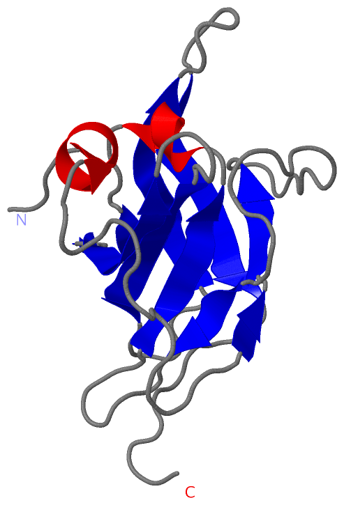
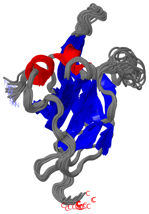
NMR Structure (1, 1)
 PROSITEUniProtKBPDB
No.IDACDescriptionIDLocationCountLocation
1NEATPS50978 NEAT domain profile.ISDH_STAAS105-232
345-471
543-660
  1A:20-144
-
-
NMR Structure * (1, 1)
 PROSITEUniProtKBPDB
No.IDACDescriptionIDLocationCountLocation
1NEATPS50978 NEAT domain profile.ISDH_STAAS105-232
345-471
543-660
  1A:20-144
-
-

(-) Exons   (0, 0)

(no "Exon" information available for 2H3K)

(-) Sequences/Alignments

NMR Structure
   Reformat: Number of residues per line =  ('0' or empty: single-line sequence representation)
  Number of residues per labelling interval =   
  UniProt sequence: complete  aligned part    
   Show mapping: SCOP domains CATH domains Pfam domains Secondary structure (by author)
SAPs(SNPs) PROSITE motifs Exons
(details for a mapped element are shown in a popup box when the mouse pointer rests over it)
Chain A from PDB  Type:PROTEIN  Length:144
 aligned with ISDH_STAAS | Q6G8J7 from UniProtKB/Swiss-Prot  Length:895

    Alignment length:144
                                    95       105       115       125       135       145       155       165       175       185       195       205       215       225    
           ISDH_STAAS    86 ADESLKDAIKDPALENKEHDIGPREQVNFQLLDKNNETQYYHFFSIKDPADVYYTKKKAEVELDINTASTWKKFEVYENNQKLPVRLVSYSPVPEDHAYIRFPVSDGTQELKIVSSTQIDDGEETNYDYTKLVFAKPIYNDPSL 229
               SCOP domains d2h3ka1 A:1-144 Iron-regulated surface determinant protein H, IsdH                                                                               SCOP domains
               CATH domains ------------------------------------------------------------------------------------------------------------------------------------------------ CATH domains
               Pfam domains ------------------------------------------------------------------------------------------------------------------------------------------------ Pfam domains
         Sec.struct. author ...hhhhhh................ee.eeee.................eee.......eeeeee.hhhhh....eee..eee..eeeeee.....eeeeeee......eeeeeeee.......eeeeeeeeee.......... Sec.struct. author
                 SAPs(SNPs) ------------------------------------------------------------------------------------------------------------------------------------------------ SAPs(SNPs)
                    PROSITE -------------------NEAT  PDB: A:20-144 UniProt: 105-232                                                                                          PROSITE
                 Transcript ------------------------------------------------------------------------------------------------------------------------------------------------ Transcript
                 2h3k A   1 ADESLKDAIKDPALENKEHDIGPREQVNFQLLDKNNETQYYHFFSIKDPADVYYTKKKAEVELDINTASTWKKFEVYENNQKLPVRLVSYSPVPEDHAYIRFPVSDGTQELKIVSSTQIDDGEETNYDYTKLVFAKPIYNDPSL 144
                                    10        20        30        40        50        60        70        80        90       100       110       120       130       140    

   Legend:   → Mismatch (orange background)
  - → Gap (green background, '-', border residues have a numbering label)
    → Modified Residue (blue background, lower-case, 'x' indicates undefined single-letter code, labelled with number + name)
  x → Chemical Group (purple background, 'x', labelled with number + name, e.g. ACE or NH2)
  extra numbering lines below/above indicate numbering irregularities and modified residue names etc., number ends below/above '|'

 Classification and Annotation

(-) SCOP Domains  (1, 1)

NMR Structure

(-) CATH Domains  (0, 0)

(no "CATH Domain" information available for 2H3K)

(-) Pfam Domains  (0, 0)

(no "Pfam Domain" information available for 2H3K)

(-) Gene Ontology  (3, 3)

NMR Structure(hide GO term definitions)
Chain A   (ISDH_STAAS | Q6G8J7)
cellular component
    GO:0005618    cell wall    The rigid or semi-rigid envelope lying outside the cell membrane of plant, fungal, most prokaryotic cells and some protozoan parasites, maintaining their shape and protecting them from osmotic lysis. In plants it is made of cellulose and, often, lignin; in fungi it is composed largely of polysaccharides; in bacteria it is composed of peptidoglycan; in protozoan parasites such as Giardia species, it's made of carbohydrates and proteins.
    GO:0005576    extracellular region    The space external to the outermost structure of a cell. For cells without external protective or external encapsulating structures this refers to space outside of the plasma membrane. This term covers the host cell environment outside an intracellular parasite.
    GO:0016020    membrane    A lipid bilayer along with all the proteins and protein complexes embedded in it an attached to it.

 Visualization

(-) Interactive Views

NMR Structure
  Complete Structure
    Jena3D(integrated viewing of ligand, site, SAP, PROSITE, SCOP information)
    WebMol | AstexViewer[tm]@PDBe
(Java Applets, require no local installation except for Java; loading may be slow)
    STRAP
(Java WebStart application, automatic local installation, requires Java; full application with system access!)
    RasMol
(require local installation)
    Molscript (VRML)
(requires installation of a VRML viewer; select preferred view via VRML and generate a mono or stereo PDF format file)
 
  Ligands, Modified Residues, Ions
(no "Ligands, Modified Residues, Ions" information available for 2h3k)
 
  Sites
(no "Sites" information available for 2h3k)
 
  Cis Peptide Bonds
(no "Cis Peptide Bonds" information available for 2h3k)
 

(-) Still Images

Jmol
  protein: cartoon or spacefill or dots and stick; nucleic acid: cartoon and stick; ligands: spacefill; active site: stick
Molscript
  protein, nucleic acid: cartoon; ligands: spacefill; active site: ball and stick

 Databases and Analysis Tools

(-) Databases

Access by PDB/NDB ID
  2h3k
    Family and Domain InformationProDom | SYSTERS
    General Structural InformationGlycoscienceDB | MMDB | NDB | OCA | PDB | PDBe | PDBj | PDBsum | PDBWiki | PQS | PROTEOPEDIA
    Orientation in MembranesOPM
    Protein SurfaceSURFACE
    Secondary StructureDSSP (structure derived) | HSSP (homology derived)
    Structural GenomicsGeneCensus
    Structural NeighboursCE | VAST
    Structure ClassificationCATH | Dali | SCOP
    Validation and Original DataBMRB Data View | BMRB Restraints Grid | EDS | PROCHECK | RECOORD | WHAT_CHECK
 
Access by UniProt ID/Accession number
  ISDH_STAAS | Q6G8J7
    Comparative Protein Structure ModelsModBase
    Genomic InformationEnsembl
    Protein-protein InteractionDIP
    Sequence, Family and Domain InformationInterPro | Pfam | SMART | UniProtKB/SwissProt
 
Access by Enzyme Classificator   (EC Number)
  (no 'Enzyme Classificator' available)
    General Enzyme InformationBRENDA | EC-PDB | Enzyme | IntEnz
    PathwayKEGG | MetaCyc
 
Access by Disease Identifier   (MIM ID)
  (no 'MIM ID' available)
    Disease InformationOMIM
 
Access by GenAge ID
  (no 'GenAge ID' available)
    Age Related InformationGenAge

(-) Analysis Tools

Access by PDB/NDB ID
    Domain InformationXDom
    Interatomic Contacts of Structural UnitsCSU
    Ligand-protein ContactsLPC
    Protein CavitiescastP
    Sequence and Secondary StructurePDBCartoon
    Structure AlignmentSTRAP(Java WebStart application, automatic local installation, requires Java; full application with system access!)
    Structure and Sequence BrowserSTING
 
Access by UniProt ID/Accession number
  ISDH_STAAS | Q6G8J7
    Protein Disorder PredictionDisEMBL | FoldIndex | GLOBPLOT (for more information see DisProt)

 Related Entries

(-) Entries Sharing at Least One Protein Chain (UniProt ID)

UniProtKB/Swiss-Prot
        ISDH_STAAS | Q6G8J73ovu 3s48

(-) Related Entries Specified in the PDB File

6759