Show PDB file:   
         Plain Text   HTML   (compressed file size)
QuickSearch:   
by PDB,NDB,UniProt,PROSITE Code or Search Term(s)  
(-)Asym./Biol. Unit - manually
(-)Asym./Biol. Unit
(-)Asym./Biol. Unit - sites
collapse expand < >
Image Asym./Biol. Unit - manually
Asym./Biol. Unit - manually  (Jmol Viewer)
Image Asym./Biol. Unit
Asym./Biol. Unit  (Jmol Viewer)
Image Asym./Biol. Unit - sites
Asym./Biol. Unit - sites  (Jmol Viewer)

(-) Description

Title :  CRYSTAL STRUCTURE OF A LUMENAL PENTAPEPTIDE REPEAT PROTEIN FROM CYANOTHECE SP 51142 AT 2.3 ANGSTROM RESOLUTION. TETRAGONAL CRYSTAL FORM
 
Authors :  M. A. Kennedy, S. Ni, G. W. Buchko, H. Robinson
Date :  13 Feb 06  (Deposition) - 07 Nov 06  (Release) - 16 Nov 11  (Revision)
Method :  X-RAY DIFFRACTION
Resolution :  2.30
Chains :  Asym./Biol. Unit :  A
Keywords :  Right-Handed Beta Helix, Unknown Function (Keyword Search: [Gene Ontology, PubMed, Web (Google))
 
Reference :  G. W. Buchko, S. Ni, H. Robinson, E. A. Welsh, H. B. Pakrasi, M. A. Kenned
Characterization Of Two Potentially Universal Turn Motifs That Shape The Repeated Five-Residues Fold - Crystal Structure Of A Lumenal Pentapeptide Repeat Protein From Cyanothece 51142
Protein Sci. V. 15 2579 2006
PubMed-ID: 17075135  |  Reference-DOI: 10.1110/PS.062407506

(-) Compounds

Molecule 1 - PENTAPEPTIDE REPEAT PROTEIN
    Atcc51142
    ChainsA
    EngineeredYES
    Expression SystemESCHERICHIA COLI
    Expression System PlasmidPET30
    Expression System StrainROSETTA DE3 PLYSS
    Expression System Taxid562
    Expression System Vector TypePLASMID
    FragmentFULL-LENGTH PENTAPEPTIDE REPEAT PROTEIN MINUS N-TERMINAL 29 RESIDUES PREDICTED TO BE A SIGNAL PEPTIDE
    Organism ScientificCYANOTHECE SP. ATCC 51142
    Organism Taxid43989

 Structural Features

(-) Chains, Units

  1
Asymmetric/Biological Unit A

Summary Information (see also Sequences/Alignments below)

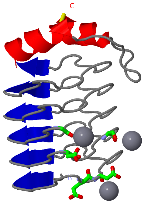
(-) Ligands, Modified Residues, Ions  (1, 3)

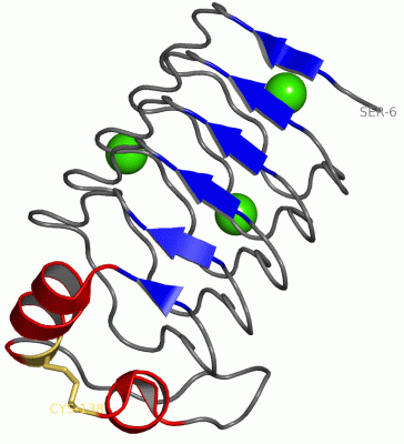
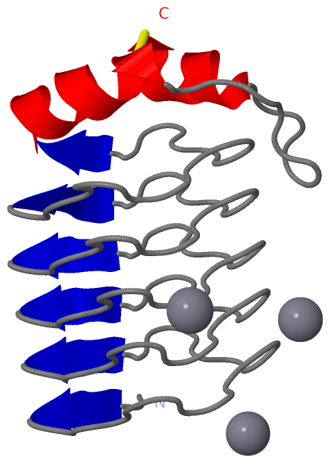
Asymmetric/Biological Unit (1, 3)
No.NameCountTypeFull Name
1CA3Ligand/IonCALCIUM ION

(-) Sites  (3, 3)

Asymmetric Unit (3, 3)
No.NameEvidenceResiduesDescription
1AC1SOFTWAREASP A:36 , GLY A:56 , HOH A:148 , HOH A:155BINDING SITE FOR RESIDUE CA A 140
2AC2SOFTWAREGLU A:17 , ASP A:18BINDING SITE FOR RESIDUE CA A 141
3AC3SOFTWARESER A:40BINDING SITE FOR RESIDUE CA A 142

(-) SS Bonds  (1, 1)

Asymmetric/Biological Unit
No.Residues
1A:118 -A:138

(-) Cis Peptide Bonds  (0, 0)

(no "Cis Peptide Bond" information available for 2G0Y)

 Sequence-Structure Mapping

(-) SAPs(SNPs)/Variants  (0, 0)

(no "SAP(SNP)/Variant" information available for 2G0Y)

(-) PROSITE Motifs  (0, 0)

(no "PROSITE Motif" information available for 2G0Y)

(-) Exons   (0, 0)

(no "Exon" information available for 2G0Y)

(-) Sequences/Alignments

Asymmetric/Biological Unit
   Reformat: Number of residues per line =  ('0' or empty: single-line sequence representation)
  Number of residues per labelling interval =   
  UniProt sequence: complete  aligned part    
   Show mapping: SCOP domains CATH domains Pfam domains Secondary structure (by author)
SAPs(SNPs) PROSITE motifs Exons
(details for a mapped element are shown in a popup box when the mouse pointer rests over it)
Chain A from PDB  Type:PROTEIN  Length:133
 aligned with RFR32_CYAA5 | B1WVN5 from UniProtKB/Swiss-Prot  Length:179

    Alignment length:133
                                    55        65        75        85        95       105       115       125       135       145       155       165       175   
          RFR32_CYAA5    46 SASYEDVKLIGEDFSGKSLTYAQFTNADLTDSNFSEADLRGAVFNGSALIGADLHGADLTNGLAYLTSFKGADLTNAVLTEAIMMRTKFDDAKITGADFSLAVLDVYEVDKLCDRADGVNPKTGVSTRESLGC 178
               SCOP domains d2g0ya_ A: Lumenal RFR-domain protein                                                                                                 SCOP domains
               CATH domains ------------------------------------------------------------------------------------------------------------------------------------- CATH domains
               Pfam domains ------------------------------------------------------------------------------------------------------------------------------------- Pfam domains
         Sec.struct. author ..eee.................eee.................eee.................eee.................eee.................eehhhhhhhhhh...........hhhhhhh. Sec.struct. author
                 SAPs(SNPs) ------------------------------------------------------------------------------------------------------------------------------------- SAPs(SNPs)
                    PROSITE ------------------------------------------------------------------------------------------------------------------------------------- PROSITE
                 Transcript ------------------------------------------------------------------------------------------------------------------------------------- Transcript
                 2g0y A   6 SASYEDVKLIGEDFSGKSLTYAQFTNADLTDSNFSEADLRGAVFNGSALIGADLHGADLTNGLAYLTSFKGADLTNAVLTEAIMMRTKFDDAKITGADFSLAVLDVYEVDKLCDRADGVNPKTGVSTRESLRC 138
                                    15        25        35        45        55        65        75        85        95       105       115       125       135   

   Legend:   → Mismatch (orange background)
  - → Gap (green background, '-', border residues have a numbering label)
    → Modified Residue (blue background, lower-case, 'x' indicates undefined single-letter code, labelled with number + name)
  x → Chemical Group (purple background, 'x', labelled with number + name, e.g. ACE or NH2)
  extra numbering lines below/above indicate numbering irregularities and modified residue names etc., number ends below/above '|'

 Classification and Annotation

(-) SCOP Domains  (1, 1)

Asymmetric/Biological Unit

(-) CATH Domains  (0, 0)

(no "CATH Domain" information available for 2G0Y)

(-) Pfam Domains  (0, 0)

(no "Pfam Domain" information available for 2G0Y)

(-) Gene Ontology  (2, 2)

Asymmetric/Biological Unit(hide GO term definitions)
Chain A   (RFR32_CYAA5 | B1WVN5)
cellular component
    GO:0009579    thylakoid    A membranous cellular structure that bears the photosynthetic pigments in plants, algae, and cyanobacteria. In cyanobacteria thylakoids are of various shapes and are attached to, or continuous with, the plasma membrane. In eukaryotes they are flattened, membrane-bounded disk-like structures located in the chloroplasts; in the chloroplasts of higher plants the thylakoids form dense stacks called grana. Isolated thylakoid preparations can carry out photosynthetic electron transport and the associated phosphorylation.
    GO:0031977    thylakoid lumen    The volume enclosed by a thylakoid membrane.

 Visualization

(-) Interactive Views

Asymmetric/Biological Unit
  Complete Structure
    Jena3D(integrated viewing of ligand, site, SAP, PROSITE, SCOP information)
    WebMol | AstexViewer[tm]@PDBe
(Java Applets, require no local installation except for Java; loading may be slow)
    STRAP
(Java WebStart application, automatic local installation, requires Java; full application with system access!)
    RasMol
(require local installation)
    Molscript (VRML)
(requires installation of a VRML viewer; select preferred view via VRML and generate a mono or stereo PDF format file)
 
  Ligands, Modified Residues, Ions
    CA  [ RasMol | Jena3D ]  +environment [ RasMol | Jena3D ]
 
  Sites
    AC1  [ RasMol ]  +environment [ RasMol ]
    AC2  [ RasMol ]  +environment [ RasMol ]
    AC3  [ RasMol ]  +environment [ RasMol ]
 
  Cis Peptide Bonds
(no "Cis Peptide Bonds" information available for 2g0y)
 

(-) Still Images

Jmol
  protein: cartoon or spacefill or dots and stick; nucleic acid: cartoon and stick; ligands: spacefill; active site: stick
Molscript
  protein, nucleic acid: cartoon; ligands: spacefill; active site: ball and stick

 Databases and Analysis Tools

(-) Databases

Access by PDB/NDB ID
  2g0y
    Family and Domain InformationProDom | SYSTERS
    General Structural InformationGlycoscienceDB | MMDB | NDB | OCA | PDB | PDBe | PDBj | PDBsum | PDBWiki | PQS | PROTEOPEDIA
    Orientation in MembranesOPM
    Protein SurfaceSURFACE
    Secondary StructureDSSP (structure derived) | HSSP (homology derived)
    Structural GenomicsGeneCensus
    Structural NeighboursCE | VAST
    Structure ClassificationCATH | Dali | SCOP
    Validation and Original DataBMRB Data View | BMRB Restraints Grid | EDS | PROCHECK | RECOORD | WHAT_CHECK
 
Access by UniProt ID/Accession number
  RFR32_CYAA5 | B1WVN5
    Comparative Protein Structure ModelsModBase
    Genomic InformationEnsembl
    Protein-protein InteractionDIP
    Sequence, Family and Domain InformationInterPro | Pfam | SMART | UniProtKB/SwissProt
 
Access by Enzyme Classificator   (EC Number)
  (no 'Enzyme Classificator' available)
    General Enzyme InformationBRENDA | EC-PDB | Enzyme | IntEnz
    PathwayKEGG | MetaCyc
 
Access by Disease Identifier   (MIM ID)
  (no 'MIM ID' available)
    Disease InformationOMIM
 
Access by GenAge ID
  (no 'GenAge ID' available)
    Age Related InformationGenAge

(-) Analysis Tools

Access by PDB/NDB ID
    Domain InformationXDom
    Interatomic Contacts of Structural UnitsCSU
    Ligand-protein ContactsLPC
    Protein CavitiescastP
    Sequence and Secondary StructurePDBCartoon
    Structure AlignmentSTRAP(Java WebStart application, automatic local installation, requires Java; full application with system access!)
    Structure and Sequence BrowserSTING
 
Access by UniProt ID/Accession number
  RFR32_CYAA5 | B1WVN5
    Protein Disorder PredictionDisEMBL | FoldIndex | GLOBPLOT (for more information see DisProt)

 Related Entries

(-) Entries Sharing at Least One Protein Chain (UniProt ID)

UniProtKB/Swiss-Prot
        RFR32_CYAA5 | B1WVN52f3l

(-) Related Entries Specified in the PDB File

2f3l THE SAME PROTEIN CRYSTALLIZED IN A TRIGONAL P3221 CRYSTAL FORM