|
|
|
|
 Description
|
|
Compounds
|
||||||||||||||||||||||||||||
Chains, Units
Summary Information (see also Sequences/Alignments below) |
Ligands, Modified Residues, Ions (0, 0)| (no "Ligand,Modified Residues,Ions" information available for 2FVN) |
Sites (0, 0)| (no "Site" information available for 2FVN) |
SS Bonds (1, 1)
NMR Structure
|
||||||||
Cis Peptide Bonds (0, 0)| (no "Cis Peptide Bond" information available for 2FVN) |
SAPs(SNPs)/Variants (0, 0)| (no "SAP(SNP)/Variant" information available for 2FVN) |
PROSITE Motifs (0, 0)| (no "PROSITE Motif" information available for 2FVN) |
Exons (0, 0)| (no "Exon" information available for 2FVN) |
Sequences/Alignments
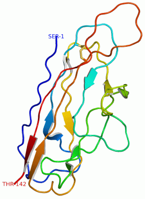
NMR StructureChain A from PDB Type:PROTEIN Length:142 aligned with AFAD_ECOLX | Q47038 from UniProtKB/Swiss-Prot Length:147 Alignment length:142 147 35 45 55 65 75 85 95 105 115 125 135 145 | - - AFAD_ECOLX 26 AAELHLESRGGSGTQLRDGAKVATGRIICREAHTGFHVWMNERQVDGRAERYVVQSKDGRHELRVRTGGDGWSPVKGEGGKGVSRPGQEEQVFFDVMADGNQDIAPGEYRFSVGGACVVPQE-------------------- - SCOP domains -d2fvna1 A:2-122 Invasin AfaD -------------------- SCOP domains CATH domains ---------------------------------------------------------------------------------------------------------------------------------------------- CATH domains Pfam domains ---------------------------------------------------------------------------------------------------------------------------------------------- Pfam domains SAPs(SNPs) ---------------------------------------------------------------------------------------------------------------------------------------------- SAPs(SNPs) PROSITE ---------------------------------------------------------------------------------------------------------------------------------------------- PROSITE Transcript ---------------------------------------------------------------------------------------------------------------------------------------------- Transcript 2fvn A 1 SAELHLESRGGSGTQLRDGAKVATGRIICREAHTGFHVWMNERQVDGRAERYVVQSKDGRHELRVRTGGDGWSPVKGEGGKGVSRPGQEEQVFFDVMADGNQDIAPGEYRFSVGGACVVPQEDNKQGFTPSGTTGTTKLTVT 142 10 20 30 40 50 60 70 80 90 100 110 120 130 140
|
||||||||||||||||||||
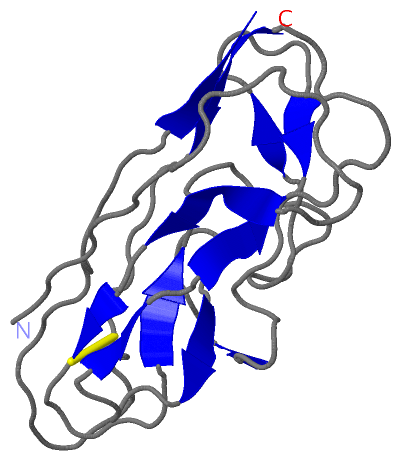
SCOP Domains (1, 1)
NMR Structure

|
CATH Domains (0, 0)| (no "CATH Domain" information available for 2FVN) |
Pfam Domains (0, 0)| (no "Pfam Domain" information available for 2FVN) |
Gene Ontology (0, 0)|
NMR Structure(hide GO term definitions)
(no "Gene Ontology" information available for 2FVN)
|
Interactive Views
|
||||||||||||||||||||||||||||||||||||||||||||||||||||||||||||||||||||||||||||||||||||||||||||||||||||||||||||||||||||
Still Images
|
||||||||||||||||
Databases
|
||||||||||||||||||||||||||||||||||||||||||||||||||||||||||||||||||||||||||||||||||||||||||||||||||||||||||||||||||||||||||||||||||||||||||||||||||||||||||||||||
Analysis Tools
|
|||||||||||||||||||||||||||||||||||||||||||||||||||||||||||||
Entries Sharing at Least One Protein Chain (UniProt ID)
Related Entries Specified in the PDB File
|
|