|
|
|
|
 Description
|
|
Compounds
|
||||||||||||||||||||||||||||||||||||||||
Chains, Units
Summary Information (see also Sequences/Alignments below) |
Ligands, Modified Residues, Ions (3, 9)
Asymmetric Unit (3, 9)
|
Sites (5, 5)
Asymmetric Unit (5, 5)
|
SS Bonds (0, 0)| (no "SS Bond" information available for 2F2E) |
Cis Peptide Bonds (1, 1)
Asymmetric Unit
|
||||||||
SAPs(SNPs)/Variants (0, 0)| (no "SAP(SNP)/Variant" information available for 2F2E) |
PROSITE Motifs (0, 0)| (no "PROSITE Motif" information available for 2F2E) |
Exons (0, 0)| (no "Exon" information available for 2F2E) |
Sequences/Alignments
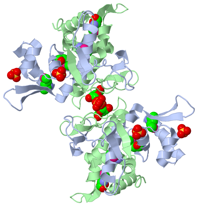
Asymmetric UnitChain A from PDB Type:PROTEIN Length:142 aligned with Q9I3B4_PSEAE | Q9I3B4 from UniProtKB/TrEMBL Length:146 Alignment length:142 14 24 34 44 54 64 74 84 94 104 114 124 134 144 Q9I3B4_PSEAE 5 TSHKQASCPVARPLDVIGDGWSMLIVRDAFEGLTRFGEFQKSLGLAKNILAARLRNLVEHGVMVAVPAESGSHQEYRLTDKGRALFPLLVAIRQWGEDYFFAPDESHVRLVERDSGQPVPRLQVRAGDGSPLAAEDTRVSRD 146 SCOP domains d2f2ea1 A:5-146 Hypothetical protein PA1607 SCOP domains CATH domains ---------2f2eA01 A:14-104 'winged helix' repressor DNA binding domain ------------------------------------------ CATH domains Pfam domains ---------------------------------------------------------------------------------------------------------------------------------------------- Pfam domains SAPs(SNPs) ---------------------------------------------------------------------------------------------------------------------------------------------- SAPs(SNPs) PROSITE ---------------------------------------------------------------------------------------------------------------------------------------------- PROSITE Transcript ---------------------------------------------------------------------------------------------------------------------------------------------- Transcript 2f2e A 5 TSHKQASCPVARPLDVIGDGWSmLIVRDAFEGLTRFGEFQKSLGLAKNILAARLRNLVEHGVmVAVPAESGSHQEYRLTDKGRALFPLLVAIRQWGEDYFFAPDESHVRLVERDSGQPVPRLQVRAGDGSPLAAEDTRVSRD 146 14 24 | 34 44 54 64 | 74 84 94 104 114 124 134 144 27-MSE 67-MSE Chain B from PDB Type:PROTEIN Length:140 aligned with Q9I3B4_PSEAE | Q9I3B4 from UniProtKB/TrEMBL Length:146 Alignment length:140 15 25 35 45 55 65 75 85 95 105 115 125 135 145 Q9I3B4_PSEAE 6 SHKQASCPVARPLDVIGDGWSMLIVRDAFEGLTRFGEFQKSLGLAKNILAARLRNLVEHGVMVAVPAESGSHQEYRLTDKGRALFPLLVAIRQWGEDYFFAPDESHVRLVERDSGQPVPRLQVRAGDGSPLAAEDTRVSR 145 SCOP domains d2f2eb_ B: Hypothetical protein PA1607 SCOP domains CATH domains --------2f2eB01 B:14-104 'winged helix' repressor DNA binding domain ----------------------------------------- CATH domains Pfam domains -------------------------------------------------------------------------------------------------------------------------------------------- Pfam domains SAPs(SNPs) -------------------------------------------------------------------------------------------------------------------------------------------- SAPs(SNPs) PROSITE -------------------------------------------------------------------------------------------------------------------------------------------- PROSITE Transcript -------------------------------------------------------------------------------------------------------------------------------------------- Transcript 2f2e B 6 SHKQASCPVARPLDVIGDGWSmLIVRDAFEGLTRFGEFQKSLGLAKNILAARLRNLVEHGVmVAVPAESGSHQEYRLTDKGRALFPLLVAIRQWGEDYFFAPDESHVRLVERDSGQPVPRLQVRAGDGSPLAAEDTRVSR 145 15 25 | 35 45 55 65 | 75 85 95 105 115 125 135 145 27-MSE 67-MSE
|
||||||||||||||||||||
SCOP Domains (1, 2)
Asymmetric Unit
 |
CATH Domains (1, 2)
Asymmetric Unit
|
Pfam Domains (0, 0)| (no "Pfam Domain" information available for 2F2E) |
Gene Ontology (0, 0)|
Asymmetric Unit(hide GO term definitions)
(no "Gene Ontology" information available for 2F2E)
|
Interactive Views
|
|||||||||||||||||||||||||||||||||||||||||||||||||||||||||||||||||||||||||||||||||||||||||||||||||||||||||||||||||||||||||||||||||||||||||||||||||||||||||||||||||||||||||||||||||||||||||||||
Still Images
|
||||||||||||||||
Databases
|
||||||||||||||||||||||||||||||||||||||||||||||||||||||||||||||||||||||||||||||||||||||||||||||||||||||||||||||||||||||||||||||||||||||||||||||||||||||||||||||||
Analysis Tools
|
|||||||||||||||||||||||||||||||||||||||||||||||||||||||||||||
Entries Sharing at Least One Protein Chain (UniProt ID)
Related Entries Specified in the PDB File
|
|