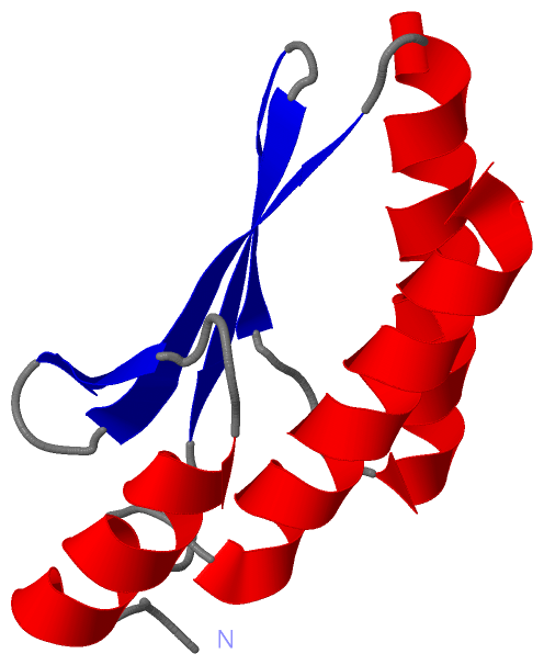
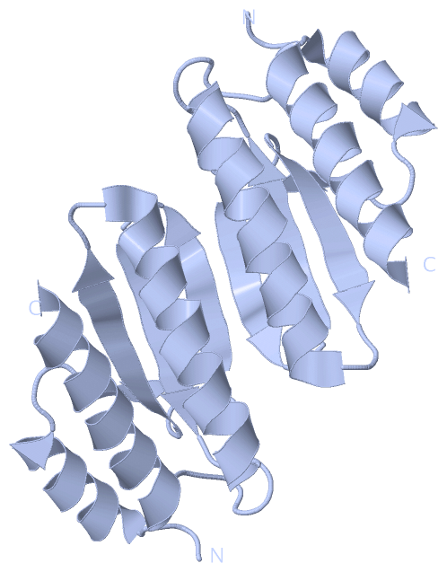
Show PDB file:   
         Plain Text   HTML   (compressed file size)
QuickSearch:   
by PDB,NDB,UniProt,PROSITE Code or Search Term(s)  
(-)Biol.Unit 1 - manually
(-)Asymmetric Unit
(-)Biological Unit 1
collapse expand < >
Image Biol.Unit 1 - manually
Biol.Unit 1 - manually  (Jmol Viewer)
Image Asymmetric Unit
Asymmetric Unit  (Jmol Viewer)
Image Biological Unit 1
Biological Unit 1  (Jmol Viewer)

(-) Description

Title :  CRYSTAL STRUCTURE OF PTERIN-4-ALPHA-CARBINOLAMINE DEHYDRATASE (PTERIN CARBINOLAMINE DEHYDRATASE) FROM GEOBACILLUS KAUSTOPHILUS HTA426
 
Authors :  T. S. Kumarevel, P. Karthe, S. Kuramitsu, S. Yokoyama, Riken Structural Genomics/Proteomics Initiative (Rsgi)
Date :  07 Feb 07  (Deposition) - 12 Feb 08  (Release) - 24 Feb 09  (Revision)
Method :  X-RAY DIFFRACTION
Resolution :  1.60
Chains :  Asym. Unit :  A
Biol. Unit 1:  A  (2x)
Keywords :  Pterin-4-Alpha-Carbinolamine Dehydratase, Coenzyme Biosyntheses, Gk1984, Structural Genomics, Nppsfa, National Project On Protein Structural And Functional Analyses, Riken Structural Genomics/Proteomics Initiative, Rsgi, Lyase (Keyword Search: [Gene Ontology, PubMed, Web (Google))
 
Reference :  T. S. Kumarevel, P. Karthe, S. Kuramitsu, S. Yokoyama
Crystal Structure Of Pterin-4-Alpha-Carbinolamine Dehydratase (Pterin Carbinolamine Dehydratase) From Geobacillus Kaustophilus Hta426
To Be Published
PubMed: search
(for further references see the PDB file header)

(-) Compounds

Molecule 1 - PTERIN-4-ALPHA-CARBINOLAMINE DEHYDRATASE
    ChainsA
    EC Number4.2.1.96
    EngineeredYES
    Expression SystemESCHERICHIA COLI
    Expression System PlasmidPET-HIS TEV
    Expression System StrainBL21 (DE3) CODONPLUS-RIL-X
    Expression System Taxid562
    Expression System Vector TypePLASMID
    GeneGK1984
    Organism ScientificGEOBACILLUS KAUSTOPHILUS
    Organism Taxid1462
    SynonymPTERIN CARBINOLAMINE DEHYDRATASE

 Structural Features

(-) Chains, Units

  1
Asymmetric Unit A
Biological Unit 1 (2x)A

Summary Information (see also Sequences/Alignments below)

(-) Ligands, Modified Residues, Ions  (0, 0)

(no "Ligand,Modified Residues,Ions" information available for 2EBB)

(-) Sites  (0, 0)

(no "Site" information available for 2EBB)

(-) SS Bonds  (0, 0)

(no "SS Bond" information available for 2EBB)

(-) Cis Peptide Bonds  (0, 0)

(no "Cis Peptide Bond" information available for 2EBB)

 Sequence-Structure Mapping

(-) SAPs(SNPs)/Variants  (0, 0)

(no "SAP(SNP)/Variant" information available for 2EBB)

(-) PROSITE Motifs  (0, 0)

(no "PROSITE Motif" information available for 2EBB)

(-) Exons   (0, 0)

(no "Exon" information available for 2EBB)

(-) Sequences/Alignments

Asymmetric Unit
   Reformat: Number of residues per line =  ('0' or empty: single-line sequence representation)
  Number of residues per labelling interval =   
  UniProt sequence: complete  aligned part    
   Show mapping: SCOP domains CATH domains Pfam domains Secondary structure (by author)
SAPs(SNPs) PROSITE motifs Exons
(details for a mapped element are shown in a popup box when the mouse pointer rests over it)
Chain A from PDB  Type:PROTEIN  Length:96
 aligned with Q5KYG7_GEOKA | Q5KYG7 from UniProtKB/TrEMBL  Length:101

    Alignment length:96
                                    10        20        30        40        50        60        70        80        90      
          Q5KYG7_GEOKA    1 MRLTEEEVQALLEKADGWKLADERWIVKKYRFQDYLQGIEFVRRIAAISENANHHPFISIDYKLITVKLSSWRAKGLTKLDFDLAKQYDEVYNQMK 96
               SCOP domains d2ebba_ A: automated matches                                                                     SCOP domains
               CATH domains ------------------------------------------------------------------------------------------------ CATH domains
               Pfam domains ------------------------------------------------------------------------------------------------ Pfam domains
         Sec.struct. author ...hhhhhhhhhhh....eee...eeeeeee..hhhhhhhhhhhhhhhhhhh....eeeee..eeeeee........hhhhhhhhhhhhhhhhhhh Sec.struct. author
                 SAPs(SNPs) ------------------------------------------------------------------------------------------------ SAPs(SNPs)
                    PROSITE ------------------------------------------------------------------------------------------------ PROSITE
                 Transcript ------------------------------------------------------------------------------------------------ Transcript
                  2ebb A  3 MRLTEEEVQALLEKADGWKLADERWIVKKYRFQDYLQGIEFVRRIAAISENANHHPFISIDYKLITVKLSSWRAKGLTKLDFDLAKQYDEVYNQMK 98
                                    12        22        32        42        52        62        72        82        92      

   Legend:   → Mismatch (orange background)
  - → Gap (green background, '-', border residues have a numbering label)
    → Modified Residue (blue background, lower-case, 'x' indicates undefined single-letter code, labelled with number + name)
  x → Chemical Group (purple background, 'x', labelled with number + name, e.g. ACE or NH2)
  extra numbering lines below/above indicate numbering irregularities and modified residue names etc., number ends below/above '|'

 Classification and Annotation

(-) SCOP Domains  (1, 1)

Asymmetric Unit

(-) CATH Domains  (0, 0)

(no "CATH Domain" information available for 2EBB)

(-) Pfam Domains  (0, 0)

(no "Pfam Domain" information available for 2EBB)

(-) Gene Ontology  (3, 3)

Asymmetric Unit(hide GO term definitions)
Chain A   (Q5KYG7_GEOKA | Q5KYG7)
molecular function
    GO:0008124    4-alpha-hydroxytetrahydrobiopterin dehydratase activity    Catalysis of the reaction: (6R)-6-(L-erythro-1,2-dihydroxypropyl)-5,6,7,8-tetrahydro-4a-hydroxypterin = (6R)-6-(L-erythro-1,2-dihydroxypropyl)-7,8-dihydro-6H-pterin + H(2)O.
    GO:0016829    lyase activity    Catalysis of the cleavage of C-C, C-O, C-N and other bonds by other means than by hydrolysis or oxidation, or conversely adding a group to a double bond. They differ from other enzymes in that two substrates are involved in one reaction direction, but only one in the other direction. When acting on the single substrate, a molecule is eliminated and this generates either a new double bond or a new ring.
biological process
    GO:0006729    tetrahydrobiopterin biosynthetic process    The chemical reactions and pathways resulting in the formation of tetrahydrobiopterin, the reduced form of biopterin (2-amino-4-hydroxy-6-(1,2-dihydroxypropyl)-pteridine). It functions as a hydroxylation coenzyme, e.g. in the conversion of phenylalanine to tyrosine.

 Visualization

(-) Interactive Views

Asymmetric Unit
  Complete Structure
    Jena3D(integrated viewing of ligand, site, SAP, PROSITE, SCOP information)
    WebMol | AstexViewer[tm]@PDBe
(Java Applets, require no local installation except for Java; loading may be slow)
    STRAP
(Java WebStart application, automatic local installation, requires Java; full application with system access!)
    RasMol
(require local installation)
    Molscript (VRML)
(requires installation of a VRML viewer; select preferred view via VRML and generate a mono or stereo PDF format file)
 
  Ligands, Modified Residues, Ions
(no "Ligands, Modified Residues, Ions" information available for 2ebb)
 
  Sites
(no "Sites" information available for 2ebb)
 
  Cis Peptide Bonds
(no "Cis Peptide Bonds" information available for 2ebb)
 
Biological Unit
  Complete Structure
    Biological Unit 1  [ Jena3D ]

(-) Still Images

Jmol
  protein: cartoon or spacefill or dots and stick; nucleic acid: cartoon and stick; ligands: spacefill; active site: stick
Molscript
  protein, nucleic acid: cartoon; ligands: spacefill; active site: ball and stick

 Databases and Analysis Tools

(-) Databases

Access by PDB/NDB ID
  2ebb
    Family and Domain InformationProDom | SYSTERS
    General Structural InformationGlycoscienceDB | MMDB | NDB | OCA | PDB | PDBe | PDBj | PDBsum | PDBWiki | PQS | PROTEOPEDIA
    Orientation in MembranesOPM
    Protein SurfaceSURFACE
    Secondary StructureDSSP (structure derived) | HSSP (homology derived)
    Structural GenomicsGeneCensus
    Structural NeighboursCE | VAST
    Structure ClassificationCATH | Dali | SCOP
    Validation and Original DataBMRB Data View | BMRB Restraints Grid | EDS | PROCHECK | RECOORD | WHAT_CHECK
 
Access by UniProt ID/Accession number
  Q5KYG7_GEOKA | Q5KYG7
    Comparative Protein Structure ModelsModBase
    Genomic InformationEnsembl
    Protein-protein InteractionDIP
    Sequence, Family and Domain InformationInterPro | Pfam | SMART | UniProtKB/TrEMBL
 
Access by Enzyme Classificator   (EC Number)
  4.2.1.96
    General Enzyme InformationBRENDA | EC-PDB | Enzyme | IntEnz
    PathwayKEGG | MetaCyc
 
Access by Disease Identifier   (MIM ID)
  (no 'MIM ID' available)
    Disease InformationOMIM
 
Access by GenAge ID
  (no 'GenAge ID' available)
    Age Related InformationGenAge

(-) Analysis Tools

Access by PDB/NDB ID
    Domain InformationXDom
    Interatomic Contacts of Structural UnitsCSU
    Ligand-protein ContactsLPC
    Protein CavitiescastP
    Sequence and Secondary StructurePDBCartoon
    Structure AlignmentSTRAP(Java WebStart application, automatic local installation, requires Java; full application with system access!)
    Structure and Sequence BrowserSTING
 
Access by UniProt ID/Accession number
  Q5KYG7_GEOKA | Q5KYG7
    Protein Disorder PredictionDisEMBL | FoldIndex | GLOBPLOT (for more information see DisProt)

 Related Entries

(-) Entries Sharing at Least One Protein Chain (UniProt ID)

(no "Entries Sharing at Least One Protein Chain" available for 2EBB)

(-) Related Entries Specified in the PDB File

(no "Related Entries Specified in the PDB File" available for 2EBB)