Show PDB file:   
         Plain Text   HTML   (compressed file size)
QuickSearch:   
by PDB,NDB,UniProt,PROSITE Code or Search Term(s)  
(-)NMR Structure - model 1
(-)NMR Structure - all models
collapse expand < >
Image NMR Structure - model 1
NMR Structure - model 1  (Jmol Viewer)
Image NMR Structure - all models
NMR Structure - all models  (Jmol Viewer)

(-) Description

Title :  SOLUTION STRUCTURE OF THE 6TH IG-LIKE DOMAIN FROM HUMAN MYOSIN-BINDING PROTEIN C, FAST-TYPE
 
Authors :  X. R. Qin, T. Nagashima, F. Hayashi, S. Yokoyama, Riken Structural Genomics/Proteomics Initiative (Rsgi)
Date :  09 Jan 07  (Deposition) - 10 Jul 07  (Release) - 24 Feb 09  (Revision)
Method :  SOLUTION NMR
Resolution :  NOT APPLICABLE
Chains :  NMR Structure  :  A  (20x)
Keywords :  Ig-Like Domain, Myosin-Binding Protein C, Fast-Type, Fast Mybp-C, C-Protein, Skeletal Muscle Fast-Isoform, Structural Genomics, Nppsfa, National Project On Protein Structural And Functional Analyses, Riken Structural Genomics/Proteomics Initiative, Rsgi, Structural Protein (Keyword Search: [Gene Ontology, PubMed, Web (Google))
 
Reference :  X. R. Qin, T. Nagashima, F. Hayashi, S. Yokoyama
Solution Structure Of The 6Th Ig-Like Domain From Human Myosin-Binding Protein C, Fast-Type
To Be Published
PubMed: search
(for further references see the PDB file header)

(-) Compounds

Molecule 1 - MYOSIN-BINDING PROTEIN C, FAST-TYPE
    ChainsA
    EngineeredYES
    Expression System PlasmidP060828-20
    Expression System Vector TypePLASMID
    FragmentIG-LIKE DOMAIN
    GeneMYBPC2, MYBPCF
    Organism CommonHUMAN
    Organism ScientificHOMO SAPIENS
    Organism Taxid9606
    Other DetailsCELL FREE PROTEIN SYNTHESIS
    SynonymFAST MYBP-C, C-PROTEIN, SKELETAL MUSCLE FAST ISOFORM

 Structural Features

(-) Chains, Units

  
NMR Structure (20x)

Summary Information (see also Sequences/Alignments below)

(-) Ligands, Modified Residues, Ions  (0, 0)

(no "Ligand,Modified Residues,Ions" information available for 2E7C)

(-) Sites  (0, 0)

(no "Site" information available for 2E7C)

(-) SS Bonds  (0, 0)

(no "SS Bond" information available for 2E7C)

(-) Cis Peptide Bonds  (1, 20)
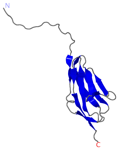
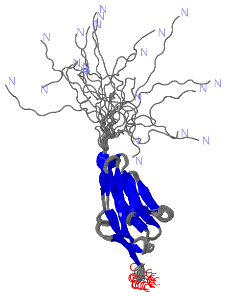
NMR Structure
No.ModelResidues
11, 2, 3, 4, 5, 6, 7, 8, 9, 10, 11, 12, 13, 14, 15, 16, 17, 18, 19, 20Lys A:51 -Pro A:52

 Sequence-Structure Mapping

(-) SAPs(SNPs)/Variants  (0, 0)

(no "SAP(SNP)/Variant" information available for 2E7C)

(-) PROSITE Motifs  (0, 0)

(no "PROSITE Motif" information available for 2E7C)

(-) Exons   (5, 5)

NMR Structure (5, 5)
 ENSEMBLUniProtKBPDB
No.Transcript IDExonExon IDGenome LocationLengthIDLocationLengthCountLocationLength
1.1ENST000003577011ENSE00001354328chr19:50936160-5093622970MYPC2_HUMAN1-770--
1.2ENST000003577012ENSE00001301795chr19:50938418-5093850790MYPC2_HUMAN7-37310--
1.3ENST000003577013ENSE00000954625chr19:50939033-5093911987MYPC2_HUMAN37-66300--
1.4ENST000003577014ENSE00000954626chr19:50939269-50939417149MYPC2_HUMAN66-115500--
1.5ENST000003577015ENSE00000954627chr19:50939874-50939991118MYPC2_HUMAN116-155400--
1.6ENST000003577016ENSE00000954628chr19:50940730-5094077849MYPC2_HUMAN155-171170--
1.7ENST000003577017ENSE00000954629chr19:50940916-5094097560MYPC2_HUMAN171-191210--
1.8ENST000003577018ENSE00000954630chr19:50944137-50944333197MYPC2_HUMAN191-257670--
1.9ENST000003577019ENSE00000954631chr19:50945438-50945570133MYPC2_HUMAN257-301450--
1.10ENST0000035770110ENSE00000954632chr19:50946751-50946875125MYPC2_HUMAN301-343430--
1.11ENST0000035770111ENSE00000954633chr19:50946968-50947073106MYPC2_HUMAN343-378360--
1.12ENST0000035770112ENSE00000954634chr19:50949137-50949309173MYPC2_HUMAN378-436590--
1.13ENST0000035770113ENSE00000954635chr19:50951482-50951647166MYPC2_HUMAN436-491560--
1.14ENST0000035770114ENSE00000721600chr19:50954086-50954192107MYPC2_HUMAN491-527370--
1.15ENST0000035770115ENSE00000721593chr19:50954537-5095456630MYPC2_HUMAN527-537110--
1.16ENST0000035770116ENSE00000954636chr19:50955121-50955260140MYPC2_HUMAN537-583470--
1.17ENST0000035770117ENSE00000954637chr19:50957277-50957436160MYPC2_HUMAN584-637540--
1.18ENST0000035770118ENSE00000954638chr19:50957522-50957626105MYPC2_HUMAN637-672360--
1.19ENST0000035770119ENSE00000954639chr19:50958365-50958553189MYPC2_HUMAN672-735640--
1.20ENST0000035770120ENSE00000954640chr19:50958767-50958901135MYPC2_HUMAN735-780461A:1-22
1.21ENST0000035770121ENSE00000869262chr19:50961844-50962011168MYPC2_HUMAN780-836571A:2-20 (gaps)57
1.22ENST0000035770122ENSE00000954641chr19:50962175-5096226389MYPC2_HUMAN836-865301A:20-4930
1.23ENST0000035770123ENSE00000869259chr19:50962368-50962563196MYPC2_HUMAN866-931661A:50-11566
1.24ENST0000035770124ENSE00000954642chr19:50963297-50963436140MYPC2_HUMAN931-977471A:115-1184
1.25ENST0000035770125ENSE00000954643chr19:50964799-50964958160MYPC2_HUMAN978-1031540--
1.26ENST0000035770126ENSE00000954644chr19:50965157-50965293137MYPC2_HUMAN1031-1076460--
1.27ENST0000035770127ENSE00001297081chr19:50967603-50967789187MYPC2_HUMAN1077-1139630--
1.28ENST0000035770128ENSE00001646879chr19:50969452-50969583132MYPC2_HUMAN1139-114130--

(-) Sequences/Alignments

NMR Structure
   Reformat: Number of residues per line =  ('0' or empty: single-line sequence representation)
  Number of residues per labelling interval =   
  UniProt sequence: complete  aligned part    
   Show mapping: SCOP domains CATH domains Pfam domains Secondary structure (by author)
SAPs(SNPs) PROSITE motifs Exons
(details for a mapped element are shown in a popup box when the mouse pointer rests over it)
Chain A from PDB  Type:PROTEIN  Length:118
 aligned with MYPC2_HUMAN | Q14324 from UniProtKB/Swiss-Prot  Length:1141

    Alignment length:156
                                   788       798       808       818       828       838       848       858       868       878       888       898       908       918       928      
          MYPC2_HUMAN   779 GSEEWVPANTEPVERCGFTVKNLPTGARILFRVVGVNIAGRSEPATLAQPVTIREIAEPPKIRLPRHLRQTYIRKVGEQLNLVVPFQGKPRPQVVWTKGGAPLDTSRVHVRTSDFDTVFFVRQAARSDSGEYELSVQIENMKDTATIRIRVVEKAG 934
               SCOP domains ------------------------------------------------------------------------------------------------------------------------------------------------------------ SCOP domains
               CATH domains ------------------------------------------------------------------------------------------------------------------------------------------------------------ CATH domains
               Pfam domains ------------------------------------------------------------------------------------------------------------------------------------------------------------ Pfam domains
         Sec.struct. author ..------------------------------------....--.............eeeee.........eeee....eeeeeeeeee...eeeeee..ee......eeee...eeeeee........eeeeeeee....ee..eeeeeee.... Sec.struct. author
                 SAPs(SNPs) ------------------------------------------------------------------------------------------------------------------------------------------------------------ SAPs(SNPs)
                    PROSITE ------------------------------------------------------------------------------------------------------------------------------------------------------------ PROSITE
           Transcript 1 (1) 1.-------------------------------------------------------Exon 1.22  PDB: A:20-49       Exon 1.23  PDB: A:50-115 UniProt: 866-931                         --- Transcript 1 (1)
           Transcript 1 (2) -Exon 1.21  PDB: A:2-20 (gaps) UniProt: 780-836           ----------------------------------------------------------------------------------------------1.24 Transcript 1 (2)
                 2e7c A   1 GS------------------------------------SGSS--GTLAQPVTIREIAEPPKIRLPRHLRQTYIRKVGEQLNLVVPFQGKPRPQVVWTKGGAPLDTSRVHVRTSDFDTVFFVRQAARSDSGEYELSVQIENMKDTATIRIRVVEKAG 118
                             |       -         -         -        |4 |  |   12        22        32        42        52        62        72        82        92       102       112      
                             2                                    3  6  7                                                                                                               

   Legend:   → Mismatch (orange background)
  - → Gap (green background, '-', border residues have a numbering label)
    → Modified Residue (blue background, lower-case, 'x' indicates undefined single-letter code, labelled with number + name)
  x → Chemical Group (purple background, 'x', labelled with number + name, e.g. ACE or NH2)
  extra numbering lines below/above indicate numbering irregularities and modified residue names etc., number ends below/above '|'

 Classification and Annotation

(-) SCOP Domains  (0, 0)

(no "SCOP Domain" information available for 2E7C)

(-) CATH Domains  (0, 0)

(no "CATH Domain" information available for 2E7C)

(-) Pfam Domains  (0, 0)

(no "Pfam Domain" information available for 2E7C)

(-) Gene Ontology  (8, 8)

NMR Structure(hide GO term definitions)
Chain A   (MYPC2_HUMAN | Q14324)
molecular function
    GO:0003779    actin binding    Interacting selectively and non-covalently with monomeric or multimeric forms of actin, including actin filaments.
    GO:0005515    protein binding    Interacting selectively and non-covalently with any protein or protein complex (a complex of two or more proteins that may include other nonprotein molecules).
    GO:0008307    structural constituent of muscle    The action of a molecule that contributes to the structural integrity of a muscle fiber.
biological process
    GO:0007155    cell adhesion    The attachment of a cell, either to another cell or to an underlying substrate such as the extracellular matrix, via cell adhesion molecules.
    GO:0006936    muscle contraction    A process in which force is generated within muscle tissue, resulting in a change in muscle geometry. Force generation involves a chemo-mechanical energy conversion step that is carried out by the actin/myosin complex activity, which generates force through ATP hydrolysis.
    GO:0030049    muscle filament sliding    The sliding of actin thin filaments and myosin thick filaments past each other in muscle contraction. This involves a process of interaction of myosin located on a thick filament with actin located on a thin filament. During this process ATP is split and forces are generated.
cellular component
    GO:0005829    cytosol    The part of the cytoplasm that does not contain organelles but which does contain other particulate matter, such as protein complexes.
    GO:0032982    myosin filament    A protein complex containing myosin heavy chains, plus associated light chains and other proteins, in which the myosin heavy chains are arranged into a filament.

 Visualization

(-) Interactive Views

NMR Structure
  Complete Structure
    Jena3D(integrated viewing of ligand, site, SAP, PROSITE, SCOP information)
    WebMol | AstexViewer[tm]@PDBe
(Java Applets, require no local installation except for Java; loading may be slow)
    STRAP
(Java WebStart application, automatic local installation, requires Java; full application with system access!)
    RasMol
(require local installation)
    Molscript (VRML)
(requires installation of a VRML viewer; select preferred view via VRML and generate a mono or stereo PDF format file)
 
  Ligands, Modified Residues, Ions
(no "Ligands, Modified Residues, Ions" information available for 2e7c)
 
  Sites
(no "Sites" information available for 2e7c)
 
  Cis Peptide Bonds
    Lys A:51 - Pro A:52   [ RasMol ]  
 

(-) Still Images

Jmol
  protein: cartoon or spacefill or dots and stick; nucleic acid: cartoon and stick; ligands: spacefill; active site: stick
Molscript
  protein, nucleic acid: cartoon; ligands: spacefill; active site: ball and stick

 Databases and Analysis Tools

(-) Databases

Access by PDB/NDB ID
  2e7c
    Family and Domain InformationProDom | SYSTERS
    General Structural InformationGlycoscienceDB | MMDB | NDB | OCA | PDB | PDBe | PDBj | PDBsum | PDBWiki | PQS | PROTEOPEDIA
    Orientation in MembranesOPM
    Protein SurfaceSURFACE
    Secondary StructureDSSP (structure derived) | HSSP (homology derived)
    Structural GenomicsGeneCensus
    Structural NeighboursCE | VAST
    Structure ClassificationCATH | Dali | SCOP
    Validation and Original DataBMRB Data View | BMRB Restraints Grid | EDS | PROCHECK | RECOORD | WHAT_CHECK
 
Access by UniProt ID/Accession number
  MYPC2_HUMAN | Q14324
    Comparative Protein Structure ModelsModBase
    Genomic InformationEnsembl
    Protein-protein InteractionDIP
    Sequence, Family and Domain InformationInterPro | Pfam | SMART | UniProtKB/SwissProt
 
Access by Enzyme Classificator   (EC Number)
  (no 'Enzyme Classificator' available)
    General Enzyme InformationBRENDA | EC-PDB | Enzyme | IntEnz
    PathwayKEGG | MetaCyc
 
Access by Disease Identifier   (MIM ID)
  (no 'MIM ID' available)
    Disease InformationOMIM
 
Access by GenAge ID
  (no 'GenAge ID' available)
    Age Related InformationGenAge

(-) Analysis Tools

Access by PDB/NDB ID
    Domain InformationXDom
    Interatomic Contacts of Structural UnitsCSU
    Ligand-protein ContactsLPC
    Protein CavitiescastP
    Sequence and Secondary StructurePDBCartoon
    Structure AlignmentSTRAP(Java WebStart application, automatic local installation, requires Java; full application with system access!)
    Structure and Sequence BrowserSTING
 
Access by UniProt ID/Accession number
  MYPC2_HUMAN | Q14324
    Protein Disorder PredictionDisEMBL | FoldIndex | GLOBPLOT (for more information see DisProt)

 Related Entries

(-) Entries Sharing at Least One Protein Chain (UniProt ID)

UniProtKB/Swiss-Prot
        MYPC2_HUMAN | Q143242edk 2edn

(-) Related Entries Specified in the PDB File

(no "Related Entries Specified in the PDB File" available for 2E7C)