|
|
|
|
 Description
|
|
Compounds
|
||||||||||||||||||||||||||||||||
Chains, Units
Summary Information (see also Sequences/Alignments below) |
Ligands, Modified Residues, Ions (2, 2)| Asymmetric Unit (2, 2) Biological Unit 1 (2, 8) |
Sites (2, 2)
Asymmetric Unit (2, 2)
|
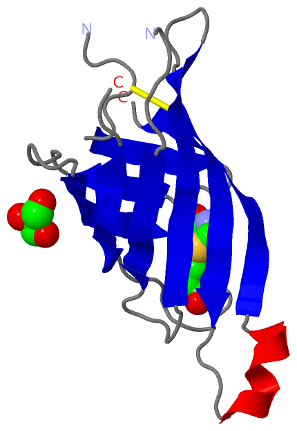
SS Bonds (1, 1)
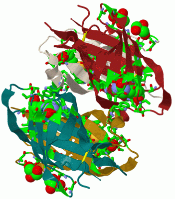
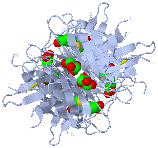
Asymmetric Unit
|
||||||||
Cis Peptide Bonds (0, 0)| (no "Cis Peptide Bond" information available for 2C1Q) |
SAPs(SNPs)/Variants (0, 0)| (no "SAP(SNP)/Variant" information available for 2C1Q) |
PROSITE Motifs (0, 0)| (no "PROSITE Motif" information available for 2C1Q) |
Exons (0, 0)| (no "Exon" information available for 2C1Q) |
Sequences/Alignments
Asymmetric Unit
Chain A from PDB Type:PROTEIN Length:123
SCOP domains d2c1qa_ A: automated matches SCOP domains
CATH domains 2c1qA00 A:1-125 [code=2.40.128.30, no name defined] CATH domains
Pfam domains --------------------------------------------------------------------------------------------------------------------------- Pfam domains
SAPs(SNPs) --------------------------------------------------------------------------------------------------------------------------- SAPs(SNPs)
PROSITE --------------------------------------------------------------------------------------------------------------------------- PROSITE
Transcript --------------------------------------------------------------------------------------------------------------------------- Transcript
2c1q A 1 TRKCELQGLWRNELGSNMTISALDVAGTFSGSYQTAVTATNKQILVSPLKGAQQPPKGQQPTFGFTVQWQFADSTTVFVGQCFVDRRGKEMLEMAWLLREEVPSRKDTWKATRVGTNVFTRVK 125
10 20 30 40 50 || 62 72 82 92 102 112 122
56|
59
|
||||||||||||||||||||
SCOP Domains (1, 1)
Asymmetric Unit
 |
CATH Domains (1, 1)
Asymmetric Unit
|
Pfam Domains (0, 0)| (no "Pfam Domain" information available for 2C1Q) |
Gene Ontology (0, 0)|
Asymmetric Unit(hide GO term definitions)
(no "Gene Ontology" information available for 2C1Q)
|
Interactive Views
|
||||||||||||||||||||||||||||||||||||||||||||||||||||||||||||||||||||||||||||||||||||||||||||||||||||||||||||||||||||||||||||||||||||||||||||||||||||||
Still Images
|
||||||||||||||||
Databases
|
||||||||||||||||||||||||||||||||||||||||||||||||||||||||||||||||||||||||||||||||||||||||||||||||||||||||||||||||||||||||||||||||||||||||||||||||||||||||||||||||
Analysis Tools
|
|||||||||||||||||||||||||||||||||||||||||||||||||||||||||||||
Entries Sharing at Least One Protein Chain (UniProt ID)
Related Entries Specified in the PDB File
|
|