|
|
|
|
 Description
|
|
Compounds
|
||||||||||||||||||||
Chains, Units
Summary Information (see also Sequences/Alignments below) |
Ligands, Modified Residues, Ions (1, 3)
Asymmetric Unit (1, 3)
|
Sites (3, 3)
Asymmetric Unit (3, 3)
|
SS Bonds (0, 0)| (no "SS Bond" information available for 2BUK) |
Cis Peptide Bonds (0, 0)| (no "Cis Peptide Bond" information available for 2BUK) |
SAPs(SNPs)/Variants (0, 0)| (no "SAP(SNP)/Variant" information available for 2BUK) |
PROSITE Motifs (0, 0)| (no "PROSITE Motif" information available for 2BUK) |
Exons (0, 0)| (no "Exon" information available for 2BUK) |
Sequences/Alignments
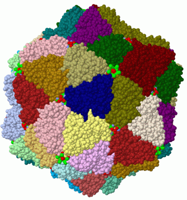
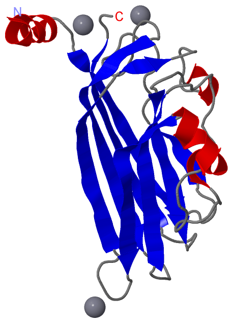
Asymmetric UnitChain A from PDB Type:PROTEIN Length:184 aligned with COAT_STNV1 | P03606 from UniProtKB/Swiss-Prot Length:196 Alignment length:184 22 32 42 52 62 72 82 92 102 112 122 132 142 152 162 172 182 192 COAT_STNV1 13 TMRAVKRMINTHLEHKRFALINSGNTNATAGTVQNLSNGIIQGDDINQRSGDQVRIVSHKLHVRGTAITVSQTFRFIWFRDNMNRGTTPTVLEVLNTANFMSQYNPITLQQKRFTILKDVTLNCSLTGESIKDRIINLPGQLVNYNGATAVAASNGPGAIFMLQIGDSLVGLWDSSYEAVYTDA 196 SCOP domains d2buka_ A: STNV coat protein SCOP domains CATH domains 2bukA00 A:12-195 [code=2.60.120.20, no name defined] CATH domains Pfam domains ---------------------------------------------------------------------------------------------------------------------------------------------------------------------------------------- Pfam domains SAPs(SNPs) ---------------------------------------------------------------------------------------------------------------------------------------------------------------------------------------- SAPs(SNPs) PROSITE ---------------------------------------------------------------------------------------------------------------------------------------------------------------------------------------- PROSITE Transcript ---------------------------------------------------------------------------------------------------------------------------------------------------------------------------------------- Transcript 2buk A 12 TMRAVKRMINTHLEHKRFALINSGNTNATAGTVQNLSNGIIQGDDINQRSGDQVRIVSHKLHVRGTAITVSQTFRFIWFRDNMNRGTTPTVLEVLNTANFMSQYNPITLQQKRFTILKDVTLNCSLTGESIKDRIINLPGQLVNYNGATAVAASNGPGAIFMLQIGDSLVGLWDSSYEAVYTDA 195 21 31 41 51 61 71 81 91 101 111 121 131 141 151 161 171 181 191
|
||||||||||||||||||||
SCOP Domains (1, 1)
Asymmetric Unit
 |
CATH Domains (1, 1)
Asymmetric Unit
|
Pfam Domains (0, 0)| (no "Pfam Domain" information available for 2BUK) |
Gene Ontology (4, 4)|
Asymmetric Unit(hide GO term definitions) Chain A (COAT_STNV1 | P03606)
|
||||||||||||||||||||||||||||||||||||
Interactive Views
|
||||||||||||||||||||||||||||||||||||||||||||||||||||||||||||||||||||||||||||||||||||||||||||||||||||||||||||||||||||||||||||||||||||||||||||||||||||||
Still Images
|
||||||||||||||||
Databases
|
||||||||||||||||||||||||||||||||||||||||||||||||||||||||||||||||||||||||||||||||||||||||||||||||||||||||||||||||||||||||||||||||||||||||||||||||||||||||||||||||
Analysis Tools
|
|||||||||||||||||||||||||||||||||||||||||||||||||||||||||||||
Entries Sharing at Least One Protein Chain (UniProt ID)
Related Entries Specified in the PDB File
|
|