|
|
|
|
 Description
|
|
Compounds
|
||||||||||||||||||||||||||||||||||||||||
Chains, Units
Summary Information (see also Sequences/Alignments below) |
Ligands, Modified Residues, Ions (4, 7)| Asymmetric/Biological Unit (4, 7) |
Sites (4, 4)
Asymmetric Unit (4, 4)
|
SS Bonds (0, 0)| (no "SS Bond" information available for 2BSW) |
Cis Peptide Bonds (1, 1)
Asymmetric/Biological Unit
|
||||||||
SAPs(SNPs)/Variants (0, 0)| (no "SAP(SNP)/Variant" information available for 2BSW) |
PROSITE Motifs (0, 0)| (no "PROSITE Motif" information available for 2BSW) |
Exons (0, 0)| (no "Exon" information available for 2BSW) |
Sequences/Alignments
Asymmetric/Biological UnitChain A from PDB Type:PROTEIN Length:145 aligned with Q65LG7_BACLD | Q65LG7 from UniProtKB/TrEMBL Length:146 Alignment length:145 11 21 31 41 51 61 71 81 91 101 111 121 131 141 Q65LG7_BACLD 2 IEVKPINAEDTYEIRHRILRPNQPLEACMYETDLLGGAFHLGGYYRGKLISIASFHKAEHSELEGEEQYQLRGMATLEGYREQKAGSTLIRHAEELLRKKGADLLWCNARTSVSGYYEKLGFSEQGEVYDIPPIGPHILMYKKLT 146 SCOP domains d2bswa1 A:2-146 Probable acetyltransferase YitI SCOP domains CATH domains ------------------------------------------------------------------------------------------------------------------------------------------------- CATH domains Pfam domains ------------------------------------------------------------------------------------------------------------------------------------------------- Pfam domains SAPs(SNPs) ------------------------------------------------------------------------------------------------------------------------------------------------- SAPs(SNPs) PROSITE ------------------------------------------------------------------------------------------------------------------------------------------------- PROSITE Transcript ------------------------------------------------------------------------------------------------------------------------------------------------- Transcript 2bsw A 2 IEVKPINAEDTYELRHRILRPNQPIEACmFESDLLRGAFHLGGYYGGKLISIASFHQAEHSELQGQKQYQLRGmATLEGYREQKAGSSLIKHAEEILRKRGADLLWCNARTSASGYYKKLGFSEQGEVFDTPPVGPHILmYKRIT 146 11 21 31 41 51 61 71 | 81 91 101 111 121 131 141 30-MSE 75-MSE 141-MSE
|
||||||||||||||||||||
SCOP Domains (1, 1)
Asymmetric/Biological Unit
 |
CATH Domains (0, 0)| (no "CATH Domain" information available for 2BSW) |
Pfam Domains (0, 0)| (no "Pfam Domain" information available for 2BSW) |
Gene Ontology (2, 2)|
Asymmetric/Biological Unit(hide GO term definitions) Chain A (Q65LG7_BACLD | Q65LG7)
|
||||||||||||||||||
Interactive Views
|
|||||||||||||||||||||||||||||||||||||||||||||||||||||||||||||||||||||||||||||||||||||||||||||||||||||||||||||||||||||||||||||||||||||||||||||||||||||||||||||||||
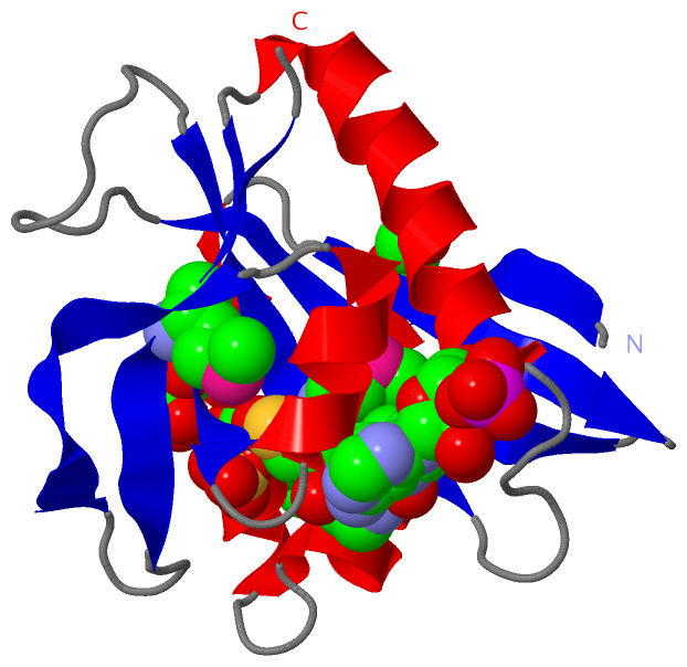
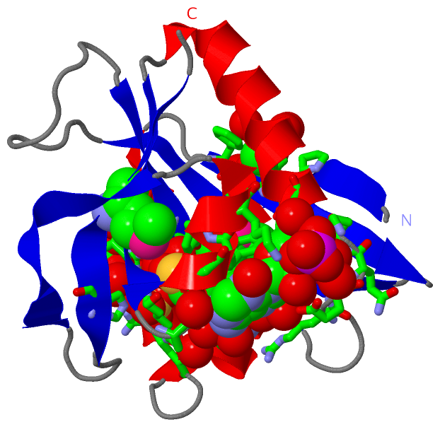
Still Images
|
||||||||||||||||
Databases
|
||||||||||||||||||||||||||||||||||||||||||||||||||||||||||||||||||||||||||||||||||||||||||||||||||||||||||||||||||||||||||||||||||||||||||||||||||||||||||||||||
Analysis Tools
|
|||||||||||||||||||||||||||||||||||||||||||||||||||||||||||||
Entries Sharing at Least One Protein Chain (UniProt ID)
Related Entries Specified in the PDB File
|
|