|
|
|
|
 Description
|
|
Compounds
|
||||||||||||||||||||||||||||||||||||
Chains, Units
Summary Information (see also Sequences/Alignments below) |
Ligands, Modified Residues, Ions (3, 6)
Asymmetric Unit (3, 6)
|
Sites (6, 6)
Asymmetric Unit (6, 6)
|
SS Bonds (0, 0)| (no "SS Bond" information available for 2BKM) |
Cis Peptide Bonds (0, 0)| (no "Cis Peptide Bond" information available for 2BKM) |
SAPs(SNPs)/Variants (0, 0)| (no "SAP(SNP)/Variant" information available for 2BKM) |
PROSITE Motifs (0, 0)| (no "PROSITE Motif" information available for 2BKM) |
Exons (0, 0)| (no "Exon" information available for 2BKM) |
Sequences/Alignments
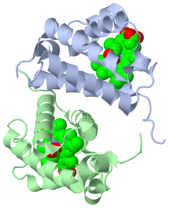
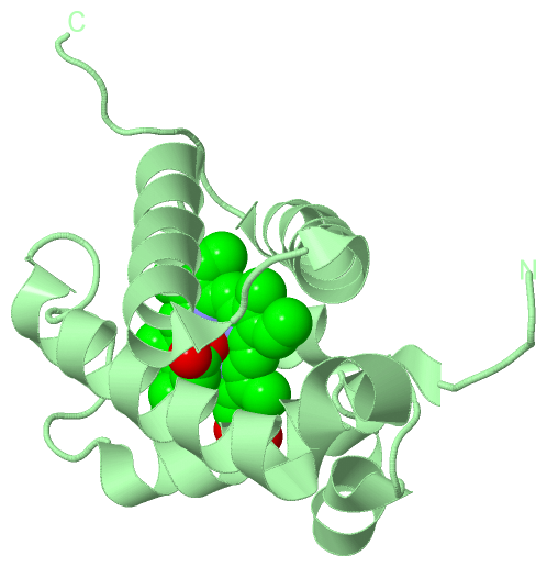
Asymmetric UnitChain A from PDB Type:PROTEIN Length:128 aligned with Q5L1S0_GEOKA | Q5L1S0 from UniProtKB/TrEMBL Length:137 Alignment length:128 12 22 32 42 52 62 72 82 92 102 112 122 Q5L1S0_GEOKA 3 EQWQTLYEAIGGEETVAKLVEAFYRRVAAHPDLRPIFPDDLTETARKQKQFLTQYLGGPPLYTAEHGHPMLRARHLRFEITPKRAEAWLACMRAAMDEIGLSGPAREQFYHRLVLTAHHMVNTPDHLD 130 SCOP domains d2bkma_ A: automated matches SCOP domains CATH domains 2bkmA00 A:3-130 Globins CATH domains Pfam domains -------------------------------------------------------------------------------------------------------------------------------- Pfam domains SAPs(SNPs) -------------------------------------------------------------------------------------------------------------------------------- SAPs(SNPs) PROSITE -------------------------------------------------------------------------------------------------------------------------------- PROSITE Transcript -------------------------------------------------------------------------------------------------------------------------------- Transcript 2bkm A 3 EQWQTLYEAIGGEETVAKLVEAFYRRVAAHPDLRPIFPDDLTETAHKQKQFLTQYLGGPPLYTAEHGHPMLRARHLRFEITPKRAEAWLACMRAAMDEIGLSGPAREQFYHRLVLTAHHMVNTPDHLD 130 12 22 32 42 52 62 72 82 92 102 112 122 Chain B from PDB Type:PROTEIN Length:128 aligned with Q5L1S0_GEOKA | Q5L1S0 from UniProtKB/TrEMBL Length:137 Alignment length:128 12 22 32 42 52 62 72 82 92 102 112 122 Q5L1S0_GEOKA 3 EQWQTLYEAIGGEETVAKLVEAFYRRVAAHPDLRPIFPDDLTETARKQKQFLTQYLGGPPLYTAEHGHPMLRARHLRFEITPKRAEAWLACMRAAMDEIGLSGPAREQFYHRLVLTAHHMVNTPDHLD 130 SCOP domains d2bkmb_ B: automated matches SCOP domains CATH domains 2bkmB00 B:3-130 Globins CATH domains Pfam domains -------------------------------------------------------------------------------------------------------------------------------- Pfam domains SAPs(SNPs) -------------------------------------------------------------------------------------------------------------------------------- SAPs(SNPs) PROSITE -------------------------------------------------------------------------------------------------------------------------------- PROSITE Transcript -------------------------------------------------------------------------------------------------------------------------------- Transcript 2bkm B 3 EQWQTLYEAIGGEETVAKLVEAFYRRVAAHPDLRPIFPDDLTETAHKQKQFLTQYLGGPPLYTAEHGHPMLRARHLRFEITPKRAEAWLACMRAAMDEIGLSGPAREQFYHRLVLTAHHMVNTPDHLD 130 12 22 32 42 52 62 72 82 92 102 112 122
|
||||||||||||||||||||
SCOP Domains (1, 2)
Asymmetric Unit
 |
CATH Domains (1, 2)
Asymmetric Unit
|
Pfam Domains (0, 0)| (no "Pfam Domain" information available for 2BKM) |
Gene Ontology (3, 3)|
Asymmetric Unit(hide GO term definitions) Chain A,B (Q5L1S0_GEOKA | Q5L1S0)
|
||||||||||||||||||||||||
Interactive Views
|
||||||||||||||||||||||||||||||||||||||||||||||||||||||||||||||||||||||||||||||||||||||||||||||||||||||||||||||||||||||||||||||||||||||||||||||||||||||||||||||||||||||||||||||||||||||||||||||
Still Images
|
||||||||||||||||
Databases
|
||||||||||||||||||||||||||||||||||||||||||||||||||||||||||||||||||||||||||||||||||||||||||||||||||||||||||||||||||||||||||||||||||||||||||||||||||||||||||||||||
Analysis Tools
|
|||||||||||||||||||||||||||||||||||||||||||||||||||||||||||||
Entries Sharing at Least One Protein Chain (UniProt ID)
Related Entries Specified in the PDB File
|
|