|
|
|
|
 Description
|
|
Compounds
|
||||||||||||||||||||||||||||||||||||||||||||||||||||||||||||
Chains, Units
Summary Information (see also Sequences/Alignments below) |
Ligands, Modified Residues, Ions (3, 8)
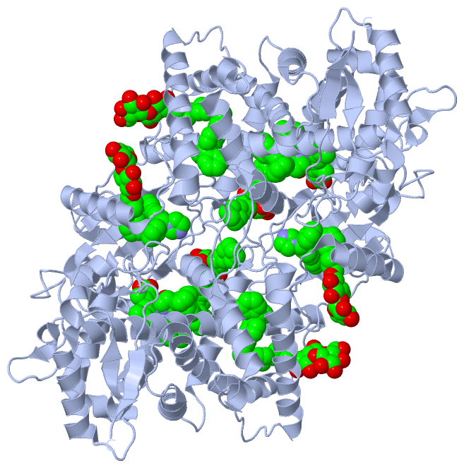
Asymmetric Unit (3, 8)
|
Sites (8, 8)
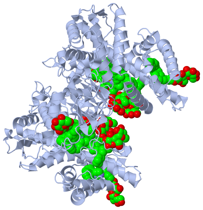
Asymmetric Unit (8, 8)
|
SS Bonds (0, 0)| (no "SS Bond" information available for 2BDM) |
Cis Peptide Bonds (1, 1)
Asymmetric Unit
|
||||||||
SAPs(SNPs)/Variants (5, 5)
Asymmetric Unit (5, 5)
|
||||||||||||||||||||||||||||||||||||||||||||||||||||||||||||||||||||||||||||||||||||||||||||||||||||||||||||||||||||||||||||||||||||||||||||||||||||||||||||||||||||||||||||||||||||||||||||||||||||||||||||||||||||||||||||||||||||||||||||||||||||||||||||||||||||||||||||||||||||||||||||||||||||||||||||||||||||||||||||||||||||||||||||||||||||||||||||||||||||||||||||||||||||||||||||||||||||||||||||||||||||||||||||||||||||||||||||||||||||||||
PROSITE Motifs (1, 1)
Asymmetric Unit (1, 1)
|
||||||||||||||||||||||||||||||||||||||||||||||||||||||||||||||||||||||||||||||||||||||||||||||||
Exons (0, 0)| (no "Exon" information available for 2BDM) |
Sequences/Alignments
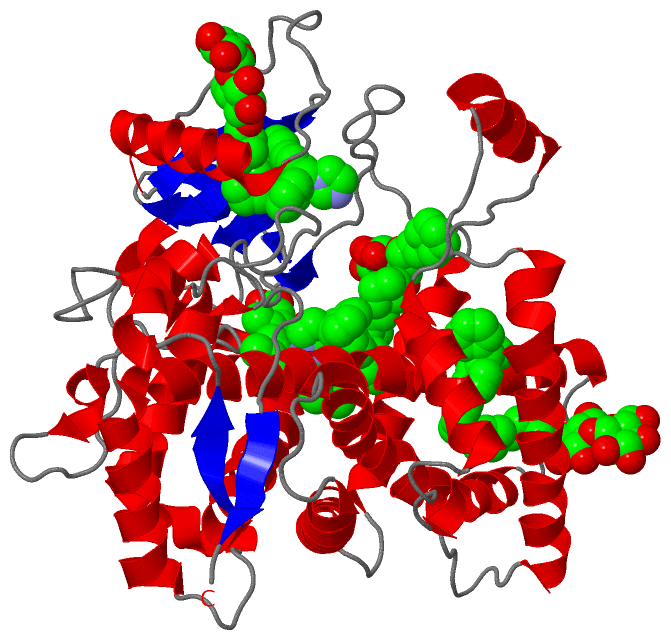
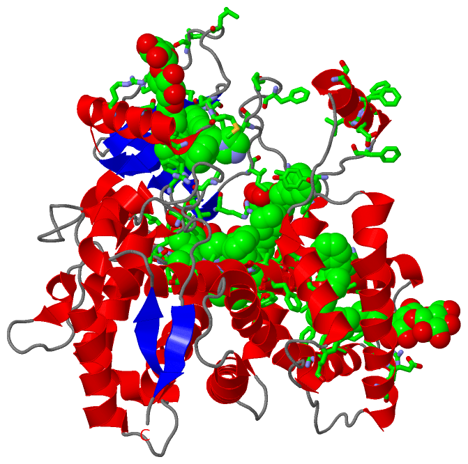
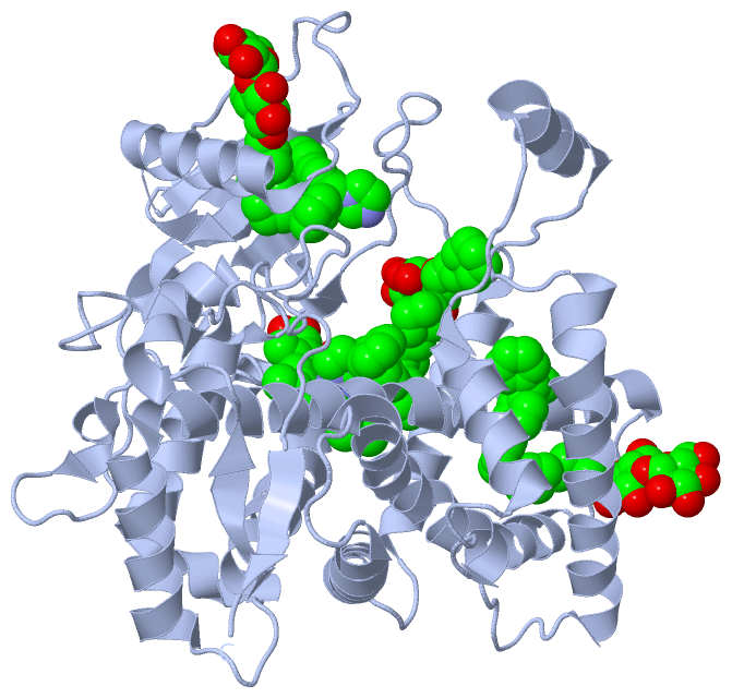
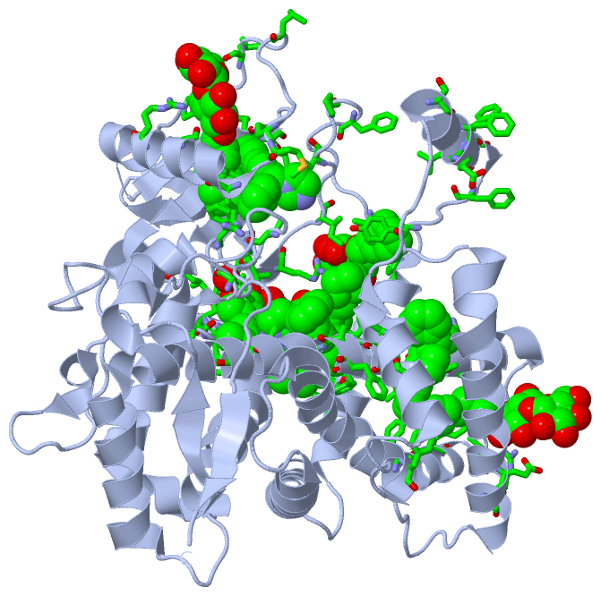
Asymmetric UnitChain A from PDB Type:PROTEIN Length:465 aligned with CP2B4_RABIT | P00178 from UniProtKB/Swiss-Prot Length:491 Alignment length:465 491 37 47 57 67 77 87 97 107 117 127 137 147 157 167 177 187 197 207 217 227 237 247 257 267 277 287 297 307 317 327 337 347 357 367 377 387 397 407 417 427 437 447 457 467 477 487 | CP2B4_RABIT 28 GRLPPGPSPLPVLGNLLQMDRKGLLRSFLRLREKYGDVFTVYLGSRPVVVLCGTDAIREALVDQAEAFSGRGKIAVVDPIFQGYGVIFANGERWRALRRFSLATMRDFGMGKRSVEERIQEEARCLVEELRKSKGALLDNTLLFHSITSNIICSIVFGKRFDYKDPVFLRLLDLFFQSFSLISSFSSQVFELFPGFLKHFPGTHRQIYRNLQEINTFIGQSVEKHRATLDPSNPRDFIDVYLLRMEKDKSDPSSEFHHQNLILTVLSLFFAGTETTSTTLRYGFLLMLKYPHVTERVQKEIEQVIGSHRPPALDDRAKMPYTDAVIHEIQRLGDLIPFGVPHTVTKDTQFRGYVIPKNTEVFPVLSSALHDPRYFETPNTFNPGHFLDANGALKRNEGFMPFSLGKRICLGEGIARTELFLFFTTILQNFSIASPVPPEDIDLTPRESGVGNVPPSYQIRFLAR- - SCOP domains d2bdma_ A: Mammalian cytochrome p450 2b4 SCOP domains CATH domains 2bdmA00 A:28-492 Cytochrome p450 CATH domains Pfam domains --------------------------------------------------------------------------------------------------------------------------------------------------------------------------------------------------------------------------------------------------------------------------------------------------------------------------------------------------------------------------------------------------------------------------------------------------------------------------------- Pfam domains SAPs(SNPs) -----------I--------------------------------------------------------------------------------------------------------------------------------------V-------------------------------------------------------------------------------------------------------------------I-----------------------L---------------------------------------------------------------------------------------------------------M------------------------------------------------------------------------ SAPs(SNPs) PROSITE -----------------------------------------------------------------------------------------------------------------------------------------------------------------------------------------------------------------------------------------------------------------------------------------------------------------------------------------------------------------------------------------------------------------CYTOCHROME------------------------------------------------------ PROSITE Transcript --------------------------------------------------------------------------------------------------------------------------------------------------------------------------------------------------------------------------------------------------------------------------------------------------------------------------------------------------------------------------------------------------------------------------------------------------------------------------------- Transcript 2bdm A 28 GKLPPGPSPLPVLGNLLQMDRKGLLRSFLRLREKYGDVFTVYLGSRPVVVLCGTDAIREALVDQAEAFSGRGKIAVVDPIFQGYGVIFANGERWRALRRFSLATMRDFGMGKRSVEERIQEEARCLVEELRKSKGALLDNTLLFHSITSNIICSIVFGKRFDYKDPVFLRLLDLFFQSFSLISSFSSQVFELFSGFLKYFPGTHRQIYRNLQEINTFIGQSVEKHRATLDPSNPRDFIDVYLLRMEKDKSDPSSEFHHQNLILTVLSLFFAGTETTSTTLRYGFLLMLKYPHVTERVQKEIEQVIGSHRPPALDDRAKMPYTDAVIHEIQRLGDLIPFGVPHTVTKDTQFRGYVIPKNTEVFPVLSSALHDPRYFETPNTFNPGHFLDANGALKRNEGFMPFSLGKRICLGEGIARTELFLFFTTILQNFSIASPVPPEDIDLTPRESGVGNVPPSYQIRFLARH 492 37 47 57 67 77 87 97 107 117 127 137 147 157 167 177 187 197 207 217 227 237 247 257 267 277 287 297 307 317 327 337 347 357 367 377 387 397 407 417 427 437 447 457 467 477 487
|
||||||||||||||||||||
SCOP Domains (1, 1)
Asymmetric Unit
 |
CATH Domains (1, 1)
Asymmetric Unit
|
Pfam Domains (0, 0)| (no "Pfam Domain" information available for 2BDM) |
Gene Ontology (14, 14)|
Asymmetric Unit(hide GO term definitions) Chain A (CP2B4_RABIT | P00178)
|
||||||||||||||||||||||||||||||||||||||||||||||||||||||||||||||||||||||||||||||||||||||||||||||||||||||
Interactive Views
|
||||||||||||||||||||||||||||||||||||||||||||||||||||||||||||||||||||||||||||||||||||||||||||||||||||||||||||||||||||||||||||||||||||||||||||||||||||||||||||||||||||||||||||||||||||||||||||||||||||||||||||||||||
Still Images
|
||||||||||||||||
Databases
|
||||||||||||||||||||||||||||||||||||||||||||||||||||||||||||||||||||||||||||||||||||||||||||||||||||||||||||||||||||||||||||||||||||||||||||||||||||||||||||||||
Analysis Tools
|
|||||||||||||||||||||||||||||||||||||||||||||||||||||||||||||
Entries Sharing at Least One Protein Chain (UniProt ID)
Related Entries Specified in the PDB File
|
|