|
|
|
|
 Description
|
|
Compounds
|
||||||||||||||||||||||||||||
Chains, Units
Summary Information (see also Sequences/Alignments below) |
Ligands, Modified Residues, Ions (3, 4)| Asymmetric Unit (3, 4) Biological Unit 1 (2, 6) |
Sites (3, 3)
Asymmetric Unit (3, 3)
|
SS Bonds (0, 0)| (no "SS Bond" information available for 2ASF) |
Cis Peptide Bonds (0, 0)| (no "Cis Peptide Bond" information available for 2ASF) |
SAPs(SNPs)/Variants (0, 0)| (no "SAP(SNP)/Variant" information available for 2ASF) |
PROSITE Motifs (0, 0)| (no "PROSITE Motif" information available for 2ASF) |
Exons (0, 0)| (no "Exon" information available for 2ASF) |
Sequences/Alignments
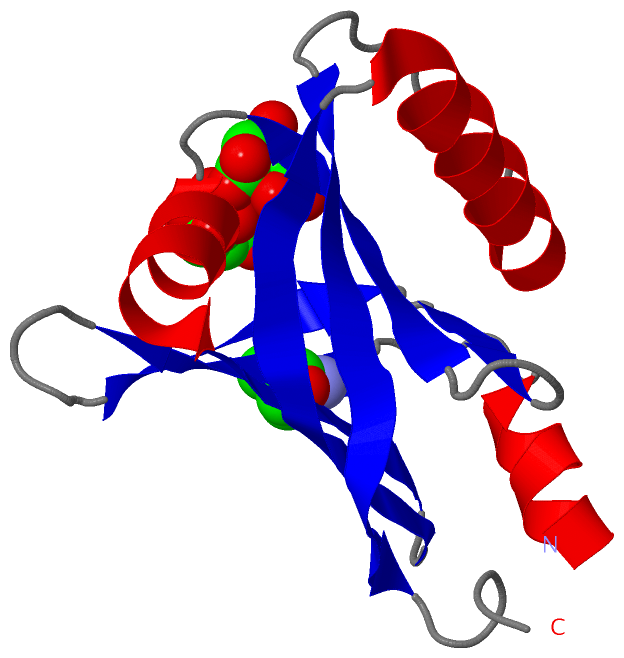
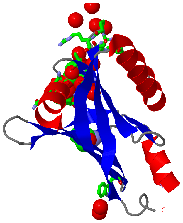
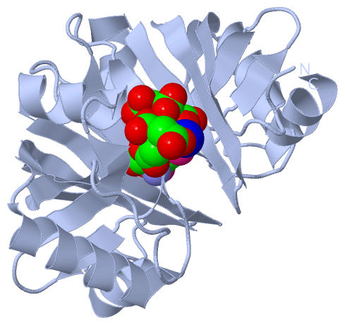
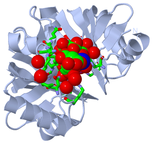
Asymmetric UnitChain A from PDB Type:PROTEIN Length:125 aligned with FBVR_MYCTO | P9WLL6 from UniProtKB/Swiss-Prot Length:137 Alignment length:125 20 30 40 50 60 70 80 90 100 110 120 130 FBVR_MYCTO 11 SDDALAFLSERHLAMLTTLRADNSPHVVAVGFTFDPKTHIARVITTGGSQKAVNADRSGLAVLSQVDGARWLSLEGRAAVNSDIDAVRDAELRYAQRYRTPRPNPRRVVIEVQIERVLGSADLLD 135 SCOP domains d2asfa1 A:11-135 Hypothetical protein Rv2074 SCOP domains CATH domains ----------------------------------------------------------------------------------------------------------------------------- CATH domains Pfam domains ----------------------------------------------------------------------------------------------------------------------------- Pfam domains SAPs(SNPs) ----------------------------------------------------------------------------------------------------------------------------- SAPs(SNPs) PROSITE ----------------------------------------------------------------------------------------------------------------------------- PROSITE Transcript ----------------------------------------------------------------------------------------------------------------------------- Transcript 2asf A 11 SDDALAFLSERHLAmLTTLRADNSPHVVAVGFTFDPKTHIARVITTGGSQKAVNADRSGLAVLSQVDGARWLSLEGRAAVNSDIDAVRDAELRYAQRYRTPRPNPRRVVIEVQIERVLGSADLLD 135 20 | 30 40 50 60 70 80 90 100 110 120 130 25-MSE Chain A from PDB Type:PROTEIN Length:125 aligned with FBVR_MYCTU | P9WLL7 from UniProtKB/Swiss-Prot Length:137 Alignment length:125 20 30 40 50 60 70 80 90 100 110 120 130 FBVR_MYCTU 11 SDDALAFLSERHLAMLTTLRADNSPHVVAVGFTFDPKTHIARVITTGGSQKAVNADRSGLAVLSQVDGARWLSLEGRAAVNSDIDAVRDAELRYAQRYRTPRPNPRRVVIEVQIERVLGSADLLD 135 SCOP domains d2asfa1 A:11-135 Hypothetical protein Rv2074 SCOP domains CATH domains ----------------------------------------------------------------------------------------------------------------------------- CATH domains Pfam domains ----------------------------------------------------------------------------------------------------------------------------- Pfam domains SAPs(SNPs) ----------------------------------------------------------------------------------------------------------------------------- SAPs(SNPs) PROSITE ----------------------------------------------------------------------------------------------------------------------------- PROSITE Transcript ----------------------------------------------------------------------------------------------------------------------------- Transcript 2asf A 11 SDDALAFLSERHLAmLTTLRADNSPHVVAVGFTFDPKTHIARVITTGGSQKAVNADRSGLAVLSQVDGARWLSLEGRAAVNSDIDAVRDAELRYAQRYRTPRPNPRRVVIEVQIERVLGSADLLD 135 20 | 30 40 50 60 70 80 90 100 110 120 130 25-MSE
|
||||||||||||||||||||
SCOP Domains (1, 1)
Asymmetric Unit

|
CATH Domains (0, 0)| (no "CATH Domain" information available for 2ASF) |
Pfam Domains (0, 0)| (no "Pfam Domain" information available for 2ASF) |
Gene Ontology (8, 13)|
Asymmetric Unit(hide GO term definitions) Chain A (FBVR_MYCTU | P9WLL7)
Chain A (FBVR_MYCTO | P9WLL6)
|
||||||||||||||||||||||||||||||||||||||||||||||||||||||||||||||||||||||||||||||||||||||||||||||||||||||||||||
Interactive Views
|
||||||||||||||||||||||||||||||||||||||||||||||||||||||||||||||||||||||||||||||||||||||||||||||||||||||||||||||||||||||||||||||||||||||||||||||||||||||||||||||||||||
Still Images
|
||||||||||||||||
Databases
|
||||||||||||||||||||||||||||||||||||||||||||||||||||||||||||||||||||||||||||||||||||||||||||||||||||||||||||||||||||||||||||||||||||||||||||||||||||||||||||||||||||||||||||||||||||||||||
Analysis Tools
|
||||||||||||||||||||||||||||||||||||||||||||||||||||||||||||||||||||||||
Entries Sharing at Least One Protein Chain (UniProt ID)
Related Entries Specified in the PDB File
|
|