Show PDB file:   
         Plain Text   HTML   (compressed file size)
QuickSearch:   
by PDB,NDB,UniProt,PROSITE Code or Search Term(s)  
(-)Asymmetric Unit
(-)Biological Unit 1
collapse expand < >
Image Asymmetric Unit
Asymmetric Unit  (Jmol Viewer)
Image Biological Unit 1
Biological Unit 1  (Jmol Viewer)

(-) Description

Title :  CRYSTAL STRUCTURES OF THE MTHK RCK DOMAIN
 
Authors :  J. Dong, N. Shi, I. Berke, L. Chen, Y. Jiang
Date :  22 Jul 05  (Deposition) - 25 Oct 05  (Release) - 13 Jul 11  (Revision)
Method :  X-RAY DIFFRACTION
Resolution :  2.80
Chains :  Asym. Unit :  A
Biol. Unit 1:  A  (2x)
Keywords :  Rossmann Fold, Helix-Turn-Helix, Metal Transport, Membrane Protein (Keyword Search: [Gene Ontology, PubMed, Web (Google))
 
Reference :  J. Dong, N. Shi, I. Berke, L. Chen, Y. Jiang
Structures Of The Mthk Rck Domain And The Effect Of Ca2+ On Gating Ring Stability
J. Biol. Chem. V. 280 41716 2005
PubMed-ID: 16227203  |  Reference-DOI: 10.1074/JBC.M508144200
(for further references see the PDB file header)

(-) Compounds

Molecule 1 - CALCIUM-GATED POTASSIUM CHANNEL MTHK
    ChainsA
    EngineeredYES
    Expression SystemESCHERICHIA COLI
    Expression System PlasmidPQE70
    Expression System StrainM15
    Expression System Taxid562
    Expression System Vector TypePLASMID
    GeneMTHK
    Organism ScientificMETHANOTHERMOBACTER THERMAUTOTROPHICUS
    Organism Taxid145262

 Structural Features

(-) Chains, Units

  1
Asymmetric Unit A
Biological Unit 1 (2x)A

Summary Information (see also Sequences/Alignments below)

(-) Ligands, Modified Residues, Ions  (0, 0)

(no "Ligand,Modified Residues,Ions" information available for 2AEM)

(-) Sites  (0, 0)

(no "Site" information available for 2AEM)

(-) SS Bonds  (0, 0)

(no "SS Bond" information available for 2AEM)

(-) Cis Peptide Bonds  (0, 0)

(no "Cis Peptide Bond" information available for 2AEM)

 Sequence-Structure Mapping

(-) SAPs(SNPs)/Variants  (0, 0)

(no "SAP(SNP)/Variant" information available for 2AEM)

(-) PROSITE Motifs  (2, 2)

Asymmetric Unit (2, 2)
 PROSITEUniProtKBPDB
No.IDACDescriptionIDLocationCountLocation
1RCK_NPS51201 RCK N-terminal domain profile.MTHK_METTH117-237  1A:117-237
2RCK_CPS51202 RCK C-terminal domain profile.MTHK_METTH252-336  1A:252-336
Biological Unit 1 (2, 4)
 PROSITEUniProtKBPDB
No.IDACDescriptionIDLocationCountLocation
1RCK_NPS51201 RCK N-terminal domain profile.MTHK_METTH117-237  2A:117-237
2RCK_CPS51202 RCK C-terminal domain profile.MTHK_METTH252-336  2A:252-336

(-) Exons   (0, 0)

(no "Exon" information available for 2AEM)

(-) Sequences/Alignments

Asymmetric Unit
   Reformat: Number of residues per line =  ('0' or empty: single-line sequence representation)
  Number of residues per labelling interval =   
  UniProt sequence: complete  aligned part    
   Show mapping: SCOP domains CATH domains Pfam domains Secondary structure (by author)
SAPs(SNPs) PROSITE motifs Exons
(details for a mapped element are shown in a popup box when the mouse pointer rests over it)
Chain A from PDB  Type:PROTEIN  Length:226
 aligned with MTHK_METTH | O27564 from UniProtKB/Swiss-Prot  Length:336

    Alignment length:226
                                                                                                                                                                                                                                                         336  
                                   122       132       142       152       162       172       182       192       202       212       222       232       242       252       262       272       282       292       302       312       322       332   |  
           MTHK_METTH   113 AKSRHVVICGWSESTLECLRELRGSEVFVLAEDENVRKKVLRSGANFVHGDPTRVSDLEKANVRGARAVIVDLESDSETIHCILGIRKIDESVRIIAEAERYENIEQLRMAGADQVISPFVISGRLMSRSIDDGYEAMFVQDVLAEESTRRMVEVPIPEGSKLEGVSVLDADIHDVTGVIIIGVGRGDELIIDPPRDYSFRAGDIILGIGKPEEIERLKNYISA--   -
               SCOP domains d2aema1 A:113-244 Potassium channel-related protein MthK                                                                            d2aema2 A:245-338 Potassium channel-related protein MthK, C-terminal domain                    SCOP domains
               CATH domains 2aemA01 A:113-230 NAD(P)-binding Rossmann-like Domain                                                                 2aemA02 A:231-258           2aemA03 A:259-338 TrkA C-terminal domain-like                                    CATH domains
               Pfam domains ---------------------------------------------------------------------------------------------------------------------------------------------------------------------------------------------------------------------------------- Pfam domains
         Sec.struct. author .....eeee..hhhhhhhhhhh....eeeee.hhhhhhhhhh...eeee....hhhhhhhh......eeee...hhhhhhhhhhhhhhhh....eeee..hhhhhhhhhhhh..eeehhhhhhhhhhhh....hhhhhhhhhhhh.....eeeeee..........hhhhhhhhhhhh.eeeeeee..eee.............eeeeeehhhhhhhhhhhhh... Sec.struct. author
                 SAPs(SNPs) ---------------------------------------------------------------------------------------------------------------------------------------------------------------------------------------------------------------------------------- SAPs(SNPs)
                    PROSITE ----RCK_N  PDB: A:117-237 UniProt: 117-237                                                                                   --------------RCK_C  PDB: A:252-336 UniProt: 252-336                                               -- PROSITE
                 Transcript ---------------------------------------------------------------------------------------------------------------------------------------------------------------------------------------------------------------------------------- Transcript
                 2aem A 113 AKSRHVVICGWSESTLECLRELRGSEVFVLAEDENVRKKVLRSGANFVHGDPTRVSDLEKANVRGARAVIVDLESDSETIHCILGIRKIDESVRIIAEAERYENIEQLRMAGADQVISPFVISGRLMSRSIDDGYEAMFVQDVLAEESTRRMVEVPIPEGSKLEGVSVLDADIHDVTGVIIIGVGRGDELIIDPPRDYSFRAGDIILGIGKPEEIERLKNYISALV 338
                                   122       132       142       152       162       172       182       192       202       212       222       232       242       252       262       272       282       292       302       312       322       332      

   Legend:   → Mismatch (orange background)
  - → Gap (green background, '-', border residues have a numbering label)
    → Modified Residue (blue background, lower-case, 'x' indicates undefined single-letter code, labelled with number + name)
  x → Chemical Group (purple background, 'x', labelled with number + name, e.g. ACE or NH2)
  extra numbering lines below/above indicate numbering irregularities and modified residue names etc., number ends below/above '|'

 Classification and Annotation

(-) SCOP Domains  (2, 2)

Asymmetric Unit

(-) CATH Domains  (3, 3)

Asymmetric Unit
(-)
Class: Alpha Beta (26913)

(-) Pfam Domains  (0, 0)

(no "Pfam Domain" information available for 2AEM)

(-) Gene Ontology  (10, 10)

Asymmetric Unit(hide GO term definitions)
Chain A   (MTHK_METTH | O27564)
molecular function
    GO:0008324    cation transmembrane transporter activity    Enables the transfer of cation from one side of the membrane to the other.
    GO:0042802    identical protein binding    Interacting selectively and non-covalently with an identical protein or proteins.
    GO:0046872    metal ion binding    Interacting selectively and non-covalently with any metal ion.
biological process
    GO:0098655    cation transmembrane transport    A process in which a cation is transported from one side of a membrane to the other by means of some agent such as a transporter or pore.
    GO:0006811    ion transport    The directed movement of charged atoms or small charged molecules into, out of or within a cell, or between cells, by means of some agent such as a transporter or pore.
    GO:0006813    potassium ion transport    The directed movement of potassium ions (K+) into, out of or within a cell, or between cells, by means of some agent such as a transporter or pore.
    GO:0006810    transport    The directed movement of substances (such as macromolecules, small molecules, ions) or cellular components (such as complexes and organelles) into, out of or within a cell, or between cells, or within a multicellular organism by means of some agent such as a transporter, pore or motor protein.
cellular component
    GO:0016021    integral component of membrane    The component of a membrane consisting of the gene products and protein complexes having at least some part of their peptide sequence embedded in the hydrophobic region of the membrane.
    GO:0016020    membrane    A lipid bilayer along with all the proteins and protein complexes embedded in it an attached to it.
    GO:0005886    plasma membrane    The membrane surrounding a cell that separates the cell from its external environment. It consists of a phospholipid bilayer and associated proteins.
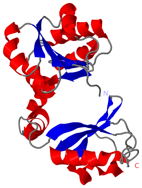
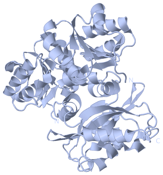
 Visualization

(-) Interactive Views

Asymmetric Unit
  Complete Structure
    Jena3D(integrated viewing of ligand, site, SAP, PROSITE, SCOP information)
    WebMol | AstexViewer[tm]@PDBe
(Java Applets, require no local installation except for Java; loading may be slow)
    STRAP
(Java WebStart application, automatic local installation, requires Java; full application with system access!)
    RasMol
(require local installation)
    Molscript (VRML)
(requires installation of a VRML viewer; select preferred view via VRML and generate a mono or stereo PDF format file)
 
  Ligands, Modified Residues, Ions
(no "Ligands, Modified Residues, Ions" information available for 2aem)
 
  Sites
(no "Sites" information available for 2aem)
 
  Cis Peptide Bonds
(no "Cis Peptide Bonds" information available for 2aem)
 
Biological Unit
  Complete Structure
    Biological Unit 1  [ Jena3D ]

(-) Still Images

Jmol
  protein: cartoon or spacefill or dots and stick; nucleic acid: cartoon and stick; ligands: spacefill; active site: stick
Molscript
  protein, nucleic acid: cartoon; ligands: spacefill; active site: ball and stick

 Databases and Analysis Tools

(-) Databases

Access by PDB/NDB ID
  2aem
    Family and Domain InformationProDom | SYSTERS
    General Structural InformationGlycoscienceDB | MMDB | NDB | OCA | PDB | PDBe | PDBj | PDBsum | PDBWiki | PQS | PROTEOPEDIA
    Orientation in MembranesOPM
    Protein SurfaceSURFACE
    Secondary StructureDSSP (structure derived) | HSSP (homology derived)
    Structural GenomicsGeneCensus
    Structural NeighboursCE | VAST
    Structure ClassificationCATH | Dali | SCOP
    Validation and Original DataBMRB Data View | BMRB Restraints Grid | EDS | PROCHECK | RECOORD | WHAT_CHECK
 
Access by UniProt ID/Accession number
  MTHK_METTH | O27564
    Comparative Protein Structure ModelsModBase
    Genomic InformationEnsembl
    Protein-protein InteractionDIP
    Sequence, Family and Domain InformationInterPro | Pfam | SMART | UniProtKB/SwissProt
 
Access by Enzyme Classificator   (EC Number)
  (no 'Enzyme Classificator' available)
    General Enzyme InformationBRENDA | EC-PDB | Enzyme | IntEnz
    PathwayKEGG | MetaCyc
 
Access by Disease Identifier   (MIM ID)
  (no 'MIM ID' available)
    Disease InformationOMIM
 
Access by GenAge ID
  (no 'GenAge ID' available)
    Age Related InformationGenAge

(-) Analysis Tools

Access by PDB/NDB ID
    Domain InformationXDom
    Interatomic Contacts of Structural UnitsCSU
    Ligand-protein ContactsLPC
    Protein CavitiescastP
    Sequence and Secondary StructurePDBCartoon
    Structure AlignmentSTRAP(Java WebStart application, automatic local installation, requires Java; full application with system access!)
    Structure and Sequence BrowserSTING
 
Access by UniProt ID/Accession number
  MTHK_METTH | O27564
    Protein Disorder PredictionDisEMBL | FoldIndex | GLOBPLOT (for more information see DisProt)

 Related Entries

(-) Entries Sharing at Least One Protein Chain (UniProt ID)

UniProtKB/Swiss-Prot
        MTHK_METTH | O275641lnq 2aef 2aej 2fy8 2ogu 3kxd 3ldc 3ldd 3lde 3ous 3r65 3rbx 3rbz 4ei2 4hyo 4hz3 4l73 4l74 4l75 4l76 4qe7 4qe9 4ro0

(-) Related Entries Specified in the PDB File

1lnq THE SAME PROTEIN WITH TRANSMEMBRANE DOMAIN
2aef THE SAME PROTEIN WITH CA2+ BOUND FORM
2aej THE SAME PROTEIN WITHOUT CA2+ BOUND FORM