Show PDB file:   
         Plain Text   HTML   (compressed file size)
QuickSearch:   
by PDB,NDB,UniProt,PROSITE Code or Search Term(s)  
(-)Asymmetric Unit
(-)Asym. Unit - sites
(-)Biological Unit 1
(-)Biol. Unit 1 - sites
collapse expand < >
Image Asymmetric Unit
Asymmetric Unit  (Jmol Viewer)
Image Asym. Unit - sites
Asym. Unit - sites  (Jmol Viewer)
Image Biological Unit 1
Biological Unit 1  (Jmol Viewer)
Image Biol. Unit 1 - sites
Biol. Unit 1 - sites  (Jmol Viewer)

(-) Description

Title :  STRUCTURAL INSIGHTS INTO DE MULTIFUNCTIONAL VP3 PROTEIN OF BIRNAVIRUSES:GOLD DERIVATIVE
 
Authors :  A. Casanas, A. Navarro, C. Ferrer-Orta, D. Gonzalez, J. F. Rodriguez, N. Verdaguer
Date :  23 Aug 07  (Deposition) - 05 Feb 08  (Release) - 13 Jul 11  (Revision)
Method :  X-RAY DIFFRACTION
Resolution :  2.40
Chains :  Asym. Unit :  A
Biol. Unit 1:  A  (2x)
Keywords :  Helix, Capsid Protein, Hydrolase, Protease, Serine Protease, Virion, Viral Protein (Keyword Search: [Gene Ontology, PubMed, Web (Google))
 
Reference :  A. Casanas, A. Navarro, C. Ferrer-Orta, D. Gonzalez, J. F. Rodriguez, N. Verdaguer
Structural Insights Into The Multifunctional Protein Vp3 Of Birnaviruses.
Structure V. 16 29 2008
PubMed-ID: 18184581  |  Reference-DOI: 10.1016/J.STR.2007.10.023

(-) Compounds

Molecule 1 - CAPSID ASSEMBLY PROTEIN VP3
    ChainsA
    EngineeredYES
    Expression SystemSPODOPTERA FRUGIPERDA
    Expression System CommonFALL ARMYWORM
    Expression System Taxid7108
    Organism CommonGUMBORO VIRUS
    Organism ScientificINFECTIOUS BURSAL DISEASE VIRUS
    Organism Taxid10995

 Structural Features

(-) Chains, Units

  1
Asymmetric Unit A
Biological Unit 1 (2x)A

Summary Information (see also Sequences/Alignments below)

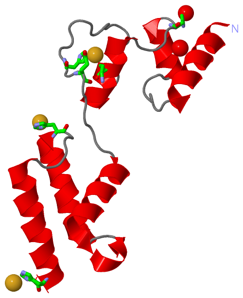
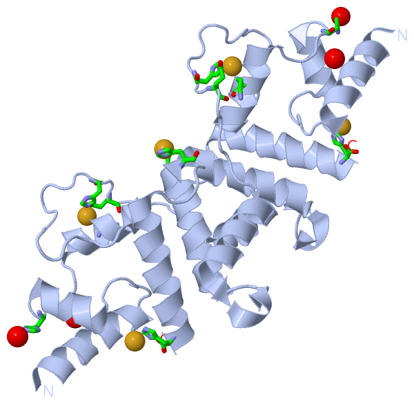
(-) Ligands, Modified Residues, Ions  (1, 3)

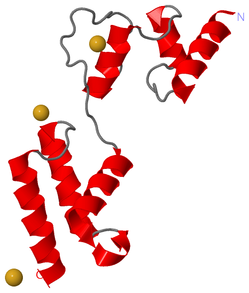
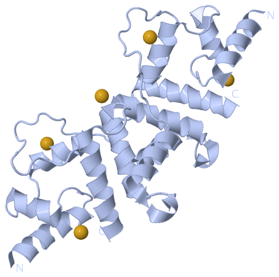
Asymmetric Unit (1, 3)
No.NameCountTypeFull Name
1AU3Ligand/IonGOLD ION
Biological Unit 1 (0, 0)
No.NameCountTypeFull Name
1AU-1Ligand/IonGOLD ION

(-) Sites  (3, 3)

Asymmetric Unit (3, 3)
No.NameEvidenceResiduesDescription
1AC1SOFTWAREHIS A:126 , LYS A:135 , HIS A:155 , GLU A:157 , HOH A:235BINDING SITE FOR RESIDUE AU A 1
2AC2SOFTWAREHIS A:220 , HOH A:247BINDING SITE FOR RESIDUE AU A 2
3AC3SOFTWAREHIS A:199BINDING SITE FOR RESIDUE AU A 3

(-) SS Bonds  (0, 0)

(no "SS Bond" information available for 2Z7J)

(-) Cis Peptide Bonds  (0, 0)

(no "Cis Peptide Bond" information available for 2Z7J)

 Sequence-Structure Mapping

(-) SAPs(SNPs)/Variants  (0, 0)

(no "SAP(SNP)/Variant" information available for 2Z7J)

(-) PROSITE Motifs  (0, 0)

(no "PROSITE Motif" information available for 2Z7J)

(-) Exons   (0, 0)

(no "Exon" information available for 2Z7J)

(-) Sequences/Alignments

Asymmetric Unit
   Reformat: Number of residues per line =  ('0' or empty: single-line sequence representation)
  Number of residues per labelling interval =   
  UniProt sequence: complete  aligned part    
   Show mapping: SCOP domains CATH domains Pfam domains Secondary structure (by author)
SAPs(SNPs) PROSITE motifs Exons
(details for a mapped element are shown in a popup box when the mouse pointer rests over it)
Chain A from PDB  Type:PROTEIN  Length:125
 aligned with POLS_IBDVP | P25220 from UniProtKB/Swiss-Prot  Length:993

    Alignment length:125
                                   840       850       860       870       880       890       900       910       920       930       940       950     
           POLS_IBDVP   831 AQREKDTRISKKMETMGIYFATPEWVALNGHRGPSPGQLKYWQNTREIPDPNEDYLDYVHAEKSRLASEEQILRAATSIYGAPGQAEPPQAFIDEVAKVYEINHGRGPNQEQMKDLLLTAMEMKH 955
               SCOP domains ----------------------------------------------------------------------------------------------------------------------------- SCOP domains
               CATH domains ----------------------------------------------------------------------------------------------------------------------------- CATH domains
               Pfam domains Birna_VP3-2z7jA01 A:96-220                                                                                                    Pfam domains
         Sec.struct. author hhhhhhhhhhhhhhh......hhhhhhhh.....hhhhhhhhhhhh.....................hhhhhhhhhhhhhhhhhhh..hhhhhhhhhhhhhhh.....hhhhhhhhhhhhhhhhh Sec.struct. author
                 SAPs(SNPs) ----------------------------------------------------------------------------------------------------------------------------- SAPs(SNPs)
                    PROSITE ----------------------------------------------------------------------------------------------------------------------------- PROSITE
                 Transcript ----------------------------------------------------------------------------------------------------------------------------- Transcript
                 2z7j A  96 AQREKDTRISKKMETMGIYFATPEWVALNGHRGPSPGQLKYWQNTREIPDPNEDYLDYVHAEKSRLASEEQILRAATSIYGAPGQAEPPQAFIDEVAKVYEINHGRGPNQEQMKDLLLTAMEMKH 220
                                   105       115       125       135       145       155       165       175       185       195       205       215     

   Legend:   → Mismatch (orange background)
  - → Gap (green background, '-', border residues have a numbering label)
    → Modified Residue (blue background, lower-case, 'x' indicates undefined single-letter code, labelled with number + name)
  x → Chemical Group (purple background, 'x', labelled with number + name, e.g. ACE or NH2)
  extra numbering lines below/above indicate numbering irregularities and modified residue names etc., number ends below/above '|'

 Classification and Annotation

(-) SCOP Domains  (0, 0)

(no "SCOP Domain" information available for 2Z7J)

(-) CATH Domains  (0, 0)

(no "CATH Domain" information available for 2Z7J)

(-) Pfam Domains  (1, 1)

Asymmetric Unit

(-) Gene Ontology  (9, 9)

Asymmetric Unit(hide GO term definitions)
Chain A   (POLS_IBDVP | P25220)
molecular function
    GO:0016787    hydrolase activity    Catalysis of the hydrolysis of various bonds, e.g. C-O, C-N, C-C, phosphoric anhydride bonds, etc. Hydrolase is the systematic name for any enzyme of EC class 3.
    GO:0046872    metal ion binding    Interacting selectively and non-covalently with any metal ion.
    GO:0008233    peptidase activity    Catalysis of the hydrolysis of a peptide bond. A peptide bond is a covalent bond formed when the carbon atom from the carboxyl group of one amino acid shares electrons with the nitrogen atom from the amino group of a second amino acid.
    GO:0008236    serine-type peptidase activity    Catalysis of the hydrolysis of peptide bonds in a polypeptide chain by a catalytic mechanism that involves a catalytic triad consisting of a serine nucleophile that is activated by a proton relay involving an acidic residue (e.g. aspartate or glutamate) and a basic residue (usually histidine).
    GO:0005198    structural molecule activity    The action of a molecule that contributes to the structural integrity of a complex or its assembly within or outside a cell.
biological process
    GO:0006508    proteolysis    The hydrolysis of proteins into smaller polypeptides and/or amino acids by cleavage of their peptide bonds.
cellular component
    GO:0030430    host cell cytoplasm    The cytoplasm of a host cell.
    GO:0019028    viral capsid    The protein coat that surrounds the infective nucleic acid in some virus particles. It comprises numerous regularly arranged subunits, or capsomeres.
    GO:0019012    virion    The complete fully infectious extracellular virus particle.

 Visualization

(-) Interactive Views

Asymmetric Unit
  Complete Structure
    Jena3D(integrated viewing of ligand, site, SAP, PROSITE, SCOP information)
    WebMol | AstexViewer[tm]@PDBe
(Java Applets, require no local installation except for Java; loading may be slow)
    STRAP
(Java WebStart application, automatic local installation, requires Java; full application with system access!)
    RasMol
(require local installation)
    Molscript (VRML)
(requires installation of a VRML viewer; select preferred view via VRML and generate a mono or stereo PDF format file)
 
  Ligands, Modified Residues, Ions
    AU  [ RasMol | Jena3D ]  +environment [ RasMol | Jena3D ]
 
  Sites
    AC1  [ RasMol ]  +environment [ RasMol ]
    AC2  [ RasMol ]  +environment [ RasMol ]
    AC3  [ RasMol ]  +environment [ RasMol ]
 
  Cis Peptide Bonds
(no "Cis Peptide Bonds" information available for 2z7j)
 
Biological Unit
  Complete Structure
    Biological Unit 1  [ Jena3D ]

(-) Still Images

Jmol
  protein: cartoon or spacefill or dots and stick; nucleic acid: cartoon and stick; ligands: spacefill; active site: stick
Molscript
  protein, nucleic acid: cartoon; ligands: spacefill; active site: ball and stick

 Databases and Analysis Tools

(-) Databases

Access by PDB/NDB ID
  2z7j
    Family and Domain InformationProDom | SYSTERS
    General Structural InformationGlycoscienceDB | MMDB | NDB | OCA | PDB | PDBe | PDBj | PDBsum | PDBWiki | PQS | PROTEOPEDIA
    Orientation in MembranesOPM
    Protein SurfaceSURFACE
    Secondary StructureDSSP (structure derived) | HSSP (homology derived)
    Structural GenomicsGeneCensus
    Structural NeighboursCE | VAST
    Structure ClassificationCATH | Dali | SCOP
    Validation and Original DataBMRB Data View | BMRB Restraints Grid | EDS | PROCHECK | RECOORD | WHAT_CHECK
 
Access by UniProt ID/Accession number
  POLS_IBDVP | P25220
    Comparative Protein Structure ModelsModBase
    Genomic InformationEnsembl
    Protein-protein InteractionDIP
    Sequence, Family and Domain InformationInterPro | Pfam | SMART | UniProtKB/SwissProt
 
Access by Enzyme Classificator   (EC Number)
  (no 'Enzyme Classificator' available)
    General Enzyme InformationBRENDA | EC-PDB | Enzyme | IntEnz
    PathwayKEGG | MetaCyc
 
Access by Disease Identifier   (MIM ID)
  (no 'MIM ID' available)
    Disease InformationOMIM
 
Access by GenAge ID
  (no 'GenAge ID' available)
    Age Related InformationGenAge

(-) Analysis Tools

Access by PDB/NDB ID
    Domain InformationXDom
    Interatomic Contacts of Structural UnitsCSU
    Ligand-protein ContactsLPC
    Protein CavitiescastP
    Sequence and Secondary StructurePDBCartoon
    Structure AlignmentSTRAP(Java WebStart application, automatic local installation, requires Java; full application with system access!)
    Structure and Sequence BrowserSTING
 
Access by UniProt ID/Accession number
  POLS_IBDVP | P25220
    Protein Disorder PredictionDisEMBL | FoldIndex | GLOBPLOT (for more information see DisProt)

 Related Entries

(-) Entries Sharing at Least One Protein Chain (UniProt ID)

UniProtKB/Swiss-Prot
        POLS_IBDVP | P252202r18

(-) Related Entries Specified in the PDB File

2r18