|
|
|
|
 Description
|
|
Compounds
|
||||||||||||||||||||||||||||||||||||
Chains, Units
Summary Information (see also Sequences/Alignments below) |
Ligands, Modified Residues, Ions (0, 0)| (no "Ligand,Modified Residues,Ions" information available for 2R18) |
Sites (0, 0)| (no "Site" information available for 2R18) |
SS Bonds (0, 0)| (no "SS Bond" information available for 2R18) |
Cis Peptide Bonds (0, 0)| (no "Cis Peptide Bond" information available for 2R18) |
SAPs(SNPs)/Variants (0, 0)| (no "SAP(SNP)/Variant" information available for 2R18) |
PROSITE Motifs (0, 0)| (no "PROSITE Motif" information available for 2R18) |
Exons (0, 0)| (no "Exon" information available for 2R18) |
Sequences/Alignments
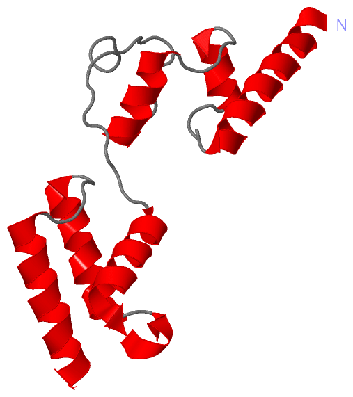
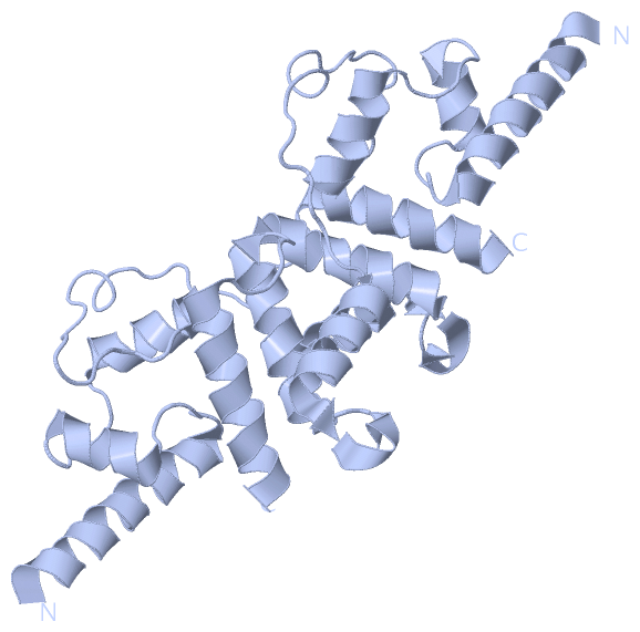
Asymmetric UnitChain A from PDB Type:PROTEIN Length:129 aligned with POLS_IBDVP | P25220 from UniProtKB/Swiss-Prot Length:993 Alignment length:129 836 846 856 866 876 886 896 906 916 926 936 946 POLS_IBDVP 827 TPEEAQREKDTRISKKMETMGIYFATPEWVALNGHRGPSPGQLKYWQNTREIPDPNEDYLDYVHAEKSRLASEEQILRAATSIYGAPGQAEPPQAFIDEVAKVYEINHGRGPNQEQMKDLLLTAMEMKH 955 SCOP domains --------------------------------------------------------------------------------------------------------------------------------- SCOP domains CATH domains --------------------------------------------------------------------------------------------------------------------------------- CATH domains Pfam domains Birna_VP3-2r18A01 A:92-220 Pfam domains SAPs(SNPs) --------------------------------------------------------------------------------------------------------------------------------- SAPs(SNPs) PROSITE --------------------------------------------------------------------------------------------------------------------------------- PROSITE Transcript --------------------------------------------------------------------------------------------------------------------------------- Transcript 2r18 A 92 TPEEAQREKDTRISKKMETMGIYFATPEWVALNGHRGPSPGQLKYWQNTREIPDPNEDYLDYVHAEKSRLASEEQILRAATSIYGAPGQAEPPQAFIDEVAKVYEINHGRGPNQEQMKDLLLTAMEMKH 220 101 111 121 131 141 151 161 171 181 191 201 211
|
||||||||||||||||||||
SCOP Domains (0, 0)| (no "SCOP Domain" information available for 2R18) |
CATH Domains (0, 0)| (no "CATH Domain" information available for 2R18) |
Pfam Domains (1, 1)
Asymmetric Unit
|
Gene Ontology (9, 9)|
Asymmetric Unit(hide GO term definitions) Chain A (POLS_IBDVP | P25220)
|
||||||||||||||||||||||||||||||||||||||||||||||||||||||||||||||||||||||||
Interactive Views
|
||||||||||||||||||||||||||||||||||||||||||||||||||||||||||||||||||||||||||||||||||||||||||||||||||||||||||||||||||||||||||||||||||||||
Still Images
|
||||||||||||||||
Databases
|
||||||||||||||||||||||||||||||||||||||||||||||||||||||||||||||||||||||||||||||||||||||||||||||||||||||||||||||||||||||||||||||||||||||||||||||||||||||||||||||||
Analysis Tools
|
|||||||||||||||||||||||||||||||||||||||||||||||||||||||||||||
Entries Sharing at Least One Protein Chain (UniProt ID)
Related Entries Specified in the PDB File
|
|