Show PDB file:   
         Plain Text   HTML   (compressed file size)
QuickSearch:   
by PDB,NDB,UniProt,PROSITE Code or Search Term(s)  
(-)Biol.Unit 1 - manually
(-)Asym.Unit - manually
(-)Asymmetric Unit
(-)Biological Unit 1
(-)Biological Unit 2
collapse expand < >
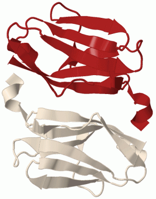
Image Biol.Unit 1 - manually
Biol.Unit 1 - manually  (Jmol Viewer)
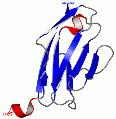
Image Asym.Unit - manually
Asym.Unit - manually  (Jmol Viewer)
Image Asymmetric Unit
Asymmetric Unit  (Jmol Viewer)
Image Biological Unit 1
Biological Unit 1  (Jmol Viewer)
Image Biological Unit 2
Biological Unit 2  (Jmol Viewer)

(-) Description

Title :  CRYSTAL STRUCTURE OF I-SET DOMAIN OF HUMAN MYOSIN BINDING PROTEINC
 
Authors :  S. Kishishita, N. Ohsawa, K. Murayama, L. Chen, Z. Liu, T. Terada, M. Shirouzu, B. Wang, S. Yokoyama, Riken Structural Genomics/Proteomics Initiative (Rsgi)
Date :  26 Apr 07  (Deposition) - 30 Oct 07  (Release) - 24 Feb 09  (Revision)
Method :  X-RAY DIFFRACTION
Resolution :  1.51
Chains :  Asym. Unit :  A
Biol. Unit 1:  A  (2x)
Biol. Unit 2:  A  (1x)
Keywords :  I-Set Domain, Myosin Binding Protein C, Cytoskeleton, Cell Adhesionn, Structural Genomics, Nppsfa, National Project On Protein Structural And Functional Analyses, Riken Structural Genomics/Proteomics Initiative, Rsgi (Keyword Search: [Gene Ontology, PubMed, Web (Google)] )
 
Reference :  S. Kishishita, N. Ohsawa, K. Murayama, T. Terada, L. Chen, Z. Liu, M. Shirouzu, B. Wang, S. Yokoyama
Crystal Structure Of I-Set Domain Of Human Myosin Binding Proteinc
To Be Published
PubMed: search
(for further references see the PDB file header)

(-) Compounds

Molecule 1 - MYOSIN-BINDING PROTEIN C, SLOW-TYPE
    Chains: A
    Engineered: YES
    Expression System Plasmid: PK050620-32
    Expression System Vector Type: PLASMID
    Fragment: I-SET DOMAIN
    Organism Common: HUMAN
    Organism Scientific: HOMO SAPIENS
    Organism Taxid: 9606
    Other Details: CELL-FREE PROTEIN SYNTHESIS
    Synonym: SLOW MYBP-C, C-PROTEIN, SKELETAL MUSCLE SLOW ISOFORM

 Structural Features

(-) Chains, Units

  1
Asymmetric Unit : A
Biological Unit 1 (2x): A
Biological Unit 2 (1x): A

Summary Information (see also Sequences/Alignments below)

(-) Ligands, Modified Residues, Ions  (0, 0)

(no "Ligand,Modified Residues,Ions" information available for 2YXM)

(-) Sites  (0, 0)

(no "Site" information available for 2YXM)

(-) SS Bonds  (0, 0)

(no "SS Bond" information available for 2YXM)

(-) Cis Peptide Bonds  (0, 0)

(no "Cis Peptide Bond" information available for 2YXM)

 Sequence-Structure Mapping

(-) SAPs(SNPs)/Variants  (0, 0)

(no "SAP(SNP)/Variant" information available for 2YXM)

(-) PROSITE Motifs  (0, 0)

(no "PROSITE Motif" information available for 2YXM)

(-) Exons   (0, 0)

(no "Exon" information available for 2YXM)

(-) Sequences/Alignments

Asymmetric Unit
   Reformat: Number of residues per line =  ('0' or empty: single-line sequence representation)
  Number of residues per labelling interval =   
  UniProt sequence: complete  aligned part    
   Show mapping: SCOP domains CATH domains Pfam domains Secondary structure (by author)
SAPs(SNPs) PROSITE motifs Exons
(details for a mapped element are shown in a popup box when the mouse pointer rests over it)
Chain A from PDB  Type:PROTEIN  Length:92
 aligned with MYPC1_HUMAN | Q00872 from UniProtKB/Swiss-Prot  Length:1141

    Alignment length:161
                                   187       197       207       217       227       237       247       257       267       277       287       297       307       317       327       337 
          MYPC1_HUMAN   178 SGEGQEDAGELDFSGLLKRREVKQQEEEPQVDVWELLKNAKPSEYEKIAFQYGITDLRGMLKRLKRMRREEKKSAAFAKILDPAYQVDKGGRVRFVVELADPKLEVKWYKNGQEIRPSTKYIFEHKGCQRILFINNCQMTDDSEYYVTAGDEKCSTELFVR 338
               SCOP domains d2y          xm                                                           a_ A: automated matches                                                                 SCOP domains
               CATH domains ----------------------------------------------------------------------------------------------------------------------------------------------------------------- CATH domains
               Pfam domains ---------------------------------------------------------------------------I-set-2yxmA01 A:9-93                                                                 - Pfam domains
         Sec.struct. author hhh----------hh-----------------------------------------------------------h.eee....eeeee....eeeeeee......eeeee..ee......eeeeee..eeeeee...hhhhheeeeeee..eeeeeeeeee Sec.struct. author
                 SAPs(SNPs) ----------------------------------------------------------------------------------------------------------------------------------------------------------------- SAPs(SNPs)
                    PROSITE ----------------------------------------------------------------------------------------------------------------------------------------------------------------- PROSITE
                 Transcript ----------------------------------------------------------------------------------------------------------------------------------------------------------------- Transcript
                 2yxm A   3 SGS----------SG-----------------------------------------------------------AAFAKILDPAYQVDKGGRVRFVVELADPKLEVKWYKNGQEIRPSTKYIFEHKGCQRILFINNCQMTDDSEYYVTAGDEKCSTELFVR  94
                              |      -   ||    -         -         -         -         -         -    |   13        23        33        43        53        63        73        83        93 
                              5          6|                                                           8                                                                                      
                                          7                                                                                                                                                  

   Legend:   → Mismatch (orange background)
  - → Gap (green background, '-', border residues have a numbering label)
    → Modified Residue (blue background, lower-case, 'x' indicates undefined single-letter code, labelled with number + name)
  x → Chemical Group (purple background, 'x', labelled with number + name, e.g. ACE or NH2)
  extra numbering lines below/above indicate numbering irregularities and modified residue names etc., number ends below/above '|'

 Classification and Annotation

(-) SCOP Domains  (1, 1)

Asymmetric Unit

(-) CATH Domains  (0, 0)

(no "CATH Domain" information available for 2YXM)

(-) Pfam Domains  (1, 1)

Asymmetric Unit
(-)
Clan: Ig (577)

(-) Gene Ontology  (11, 11)

Asymmetric Unit(hide GO term definitions)
Chain A   (MYPC1_HUMAN | Q00872)
molecular function
    GO:0003779    actin binding    Interacting selectively and non-covalently with monomeric or multimeric forms of actin, including actin filaments.
    GO:0005515    protein binding    Interacting selectively and non-covalently with any protein or protein complex (a complex of two or more proteins that may include other nonprotein molecules).
    GO:0008307    structural constituent of muscle    The action of a molecule that contributes to the structural integrity of a muscle fiber.
    GO:0031432    titin binding    Interacting selectively and non-covalently with titin, any of a family of giant proteins found in striated and smooth muscle. In striated muscle, single titin molecules span half the sarcomere, with their N- and C-termini in the Z-disc and M-line, respectively.
biological process
    GO:0007155    cell adhesion    The attachment of a cell, either to another cell or to an underlying substrate such as the extracellular matrix, via cell adhesion molecules.
    GO:0006936    muscle contraction    A process in which force is generated within muscle tissue, resulting in a change in muscle geometry. Force generation involves a chemo-mechanical energy conversion step that is carried out by the actin/myosin complex activity, which generates force through ATP hydrolysis.
    GO:0030049    muscle filament sliding    The sliding of actin thin filaments and myosin thick filaments past each other in muscle contraction. This involves a process of interaction of myosin located on a thick filament with actin located on a thin filament. During this process ATP is split and forces are generated.
    GO:1903955    positive regulation of protein targeting to mitochondrion    Any process that activates or increases the frequency, rate or extent of protein targeting to mitochondrion.
cellular component
    GO:0005829    cytosol    The part of the cytoplasm that does not contain organelles but which does contain other particulate matter, such as protein complexes.
    GO:0030016    myofibril    The contractile element of skeletal and cardiac muscle; a long, highly organized bundle of actin, myosin, and other proteins that contracts by a sliding filament mechanism.
    GO:0032982    myosin filament    A protein complex containing myosin heavy chains, plus associated light chains and other proteins, in which the myosin heavy chains are arranged into a filament.

 Visualization

(-) Interactive Views

Asymmetric Unit
  Complete Structure
    Jena3D(integrated viewing of ligand, site, SAP, PROSITE, SCOP information)
    WebMol | AstexViewer[tm]@PDBe
(Java Applets, require no local installation except for Java; loading may be slow)
    STRAP
(Java WebStart application, automatic local installation, requires Java; full application with system access!)
    RasMol
(require local installation)
    Molscript (VRML)
(requires installation of a VRML viewer; select preferred view via VRML and generate a mono or stereo PDF format file)
 
  Ligands, Modified Residues, Ions
(no "Ligands, Modified Residues, Ions" information available for 2yxm)
 
  Sites
(no "Sites" information available for 2yxm)
 
  Cis Peptide Bonds
(no "Cis Peptide Bonds" information available for 2yxm)
 
Biological Units
  Complete Structure
    Biological Unit 1  [ Jena3D ]
    Biological Unit 2  [ Jena3D ]

(-) Still Images

Jmol
  protein: cartoon or spacefill or dots and stick; nucleic acid: cartoon and stick; ligands: spacefill; active site: stick
Molscript
  protein, nucleic acid: cartoon; ligands: spacefill; active site: ball and stick

 Databases and Analysis Tools

(-) Databases

Access by PDB/NDB ID
  2yxm
    Family and Domain Information: ProDom | SYSTERS
    General Structural Information: GlycoscienceDB | MMDB | NDB | OCA | PDB | PDBe | PDBj | PDBsum | PDBWiki | PQS | PROTEOPEDIA
    Orientation in Membranes: OPM
    Protein Surface: SURFACE
    Secondary Structure: DSSP (structure derived) | HSSP (homology derived)
    Structural Genomics: GeneCensus
    Structural Neighbours: CE | VAST
    Structure Classification: CATH | Dali | SCOP
    Validation and Original Data: BMRB Data View | BMRB Restraints Grid | EDS | PROCHECK | RECOORD | WHAT_CHECK
 
Access by UniProt ID/Accession number
  MYPC1_HUMAN | Q00872
    Comparative Protein Structure Models: ModBase
    Genomic Information: Ensembl
    Protein-protein Interaction: DIP
    Sequence, Family and Domain Information: InterPro | Pfam | SMART | UniProtKB/SwissProt
 
Access by Enzyme Classificator   (EC Number)
  (no 'Enzyme Classificator' available)
    General Enzyme Information: BRENDA | EC-PDB | Enzyme | IntEnz
    Pathway: KEGG | MetaCyc
 
Access by Disease Identifier   (MIM ID)
  (no 'MIM ID' available)
    Disease Information: OMIM
 
Access by GenAge ID
  (no 'GenAge ID' available)
    Age Related Information: GenAge

(-) Analysis Tools

Access by PDB/NDB ID
    Domain Information: XDom
    Interatomic Contacts of Structural Units: CSU
    Ligand-protein Contacts: LPC
    Protein Cavities: castP
    Sequence and Secondary Structure: PDBCartoon
    Structure Alignment: STRAP(Java WebStart application, automatic local installation, requires Java; full application with system access!)
    Structure and Sequence Browser: STING
 
Access by UniProt ID/Accession number
  MYPC1_HUMAN | Q00872
    Protein Disorder Prediction: DisEMBL | FoldIndex | GLOBPLOT (for more information see DisProt)

 Related Entries

(-) Entries Sharing at Least One Protein Chain (UniProt ID)

UniProtKB/Swiss-Prot
        MYPC1_HUMAN | Q00872: 1x44 2dav 2yuv 2yuw 2yux 2yuz

(-) Related Entries Specified in the PDB File

(no "Related Entries Specified in the PDB File" available for 2YXM)