|
|
|
|
 Description
|
|
Compounds
|
||||||||||||||||||||||||||||||||||||||||
Chains, Units
Summary Information (see also Sequences/Alignments below) |
Ligands, Modified Residues, Ions (0, 0)| (no "Ligand,Modified Residues,Ions" information available for 2Y2C) |
Sites (0, 0)| (no "Site" information available for 2Y2C) |
SS Bonds (0, 0)| (no "SS Bond" information available for 2Y2C) |
Cis Peptide Bonds (6, 6)
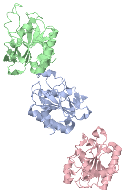
Asymmetric Unit
|
||||||||||||||||||||||||||||
SAPs(SNPs)/Variants (0, 0)| (no "SAP(SNP)/Variant" information available for 2Y2C) |
PROSITE Motifs (0, 0)| (no "PROSITE Motif" information available for 2Y2C) |
Exons (0, 0)| (no "Exon" information available for 2Y2C) |
Sequences/Alignments
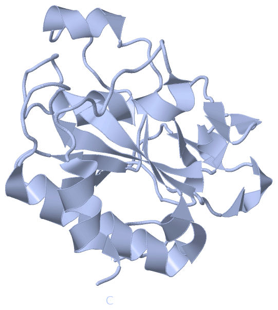
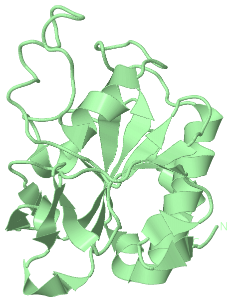
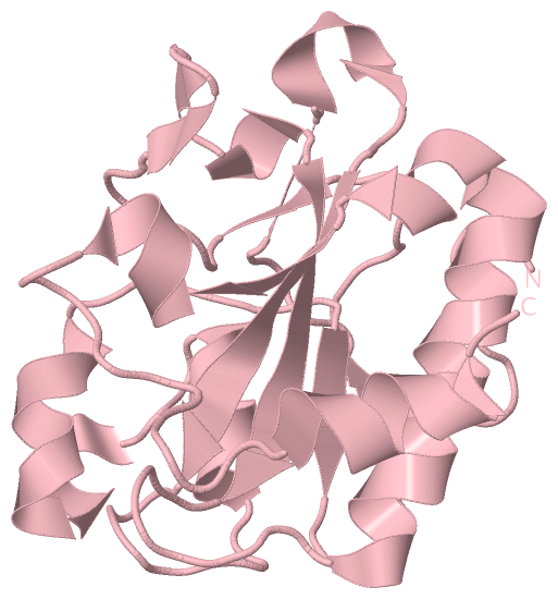
Asymmetric UnitChain A from PDB Type:PROTEIN Length:179 aligned with AMPD_CITFR | P82974 from UniProtKB/Swiss-Prot Length:187 Alignment length:179 11 21 31 41 51 61 71 81 91 101 111 121 131 141 151 161 171 AMPD_CITFR 2 LLDEGWLAEARRVPSPHYDCRPDDENPSLLVVHNISLPPGEFGGPWIDALFTGTIDPNAHPYFAGIAHLRVSAHCLIRRDGEIVQYVPFDKRAWHAGVSSYQGRERCNDFSIGIELEGTDTLAYTDAQYQQLAAVTNALITRYPAIANNMTGHCNIAPERKTDPGPSFDWARFRALVTP 180 SCOP domains d2y2ca_ A: AmpD protein SCOP domains CATH domains ----------------------------------------------------------------------------------------------------------------------------------------------------------------------------------- CATH domains Pfam domains ----------------------------------------------------------------------------------------------------------------------------------------------------------------------------------- Pfam domains SAPs(SNPs) ----------------------------------------------------------------------------------------------------------------------------------------------------------------------------------- SAPs(SNPs) PROSITE ----------------------------------------------------------------------------------------------------------------------------------------------------------------------------------- PROSITE Transcript ----------------------------------------------------------------------------------------------------------------------------------------------------------------------------------- Transcript 2y2c A 2 LLDEGWLAEARRVPSPHYDCRPDDENPSLLVVHNISLPPGEFGGPWIDALFTGTIDPNAHPYFAGIAHLRVSAHCLIRRDGEIVQYVPFDKRAWHAGVSSYQGRERCNDFSIGIELEGTDTLAYTDAQYQQLAAVTNALITRYPAIANNMTGHCNIAPERKTDPGPSFDWARFRALVTP 180 11 21 31 41 51 61 71 81 91 101 111 121 131 141 151 161 171 Chain B from PDB Type:PROTEIN Length:179 aligned with AMPD_CITFR | P82974 from UniProtKB/Swiss-Prot Length:187 Alignment length:179 10 20 30 40 50 60 70 80 90 100 110 120 130 140 150 160 170 AMPD_CITFR 1 MLLDEGWLAEARRVPSPHYDCRPDDENPSLLVVHNISLPPGEFGGPWIDALFTGTIDPNAHPYFAGIAHLRVSAHCLIRRDGEIVQYVPFDKRAWHAGVSSYQGRERCNDFSIGIELEGTDTLAYTDAQYQQLAAVTNALITRYPAIANNMTGHCNIAPERKTDPGPSFDWARFRALVT 179 SCOP domains d2y2cb_ B: AmpD protein SCOP domains CATH domains ----------------------------------------------------------------------------------------------------------------------------------------------------------------------------------- CATH domains Pfam domains ----------------------------------------------------------------------------------------------------------------------------------------------------------------------------------- Pfam domains SAPs(SNPs) ----------------------------------------------------------------------------------------------------------------------------------------------------------------------------------- SAPs(SNPs) PROSITE ----------------------------------------------------------------------------------------------------------------------------------------------------------------------------------- PROSITE Transcript ----------------------------------------------------------------------------------------------------------------------------------------------------------------------------------- Transcript 2y2c B 1 MLLDEGWLAEARRVPSPHYDCRPDDENPSLLVVHNISLPPGEFGGPWIDALFTGTIDPNAHPYFAGIAHLRVSAHCLIRRDGEIVQYVPFDKRAWHAGVSSYQGRERCNDFSIGIELEGTDTLAYTDAQYQQLAAVTNALITRYPAIANNMTGHCNIAPERKTDPGPSFDWARFRALVT 179 10 20 30 40 50 60 70 80 90 100 110 120 130 140 150 160 170 Chain C from PDB Type:PROTEIN Length:176 aligned with AMPD_CITFR | P82974 from UniProtKB/Swiss-Prot Length:187 Alignment length:176 13 23 33 43 53 63 73 83 93 103 113 123 133 143 153 163 173 AMPD_CITFR 4 DEGWLAEARRVPSPHYDCRPDDENPSLLVVHNISLPPGEFGGPWIDALFTGTIDPNAHPYFAGIAHLRVSAHCLIRRDGEIVQYVPFDKRAWHAGVSSYQGRERCNDFSIGIELEGTDTLAYTDAQYQQLAAVTNALITRYPAIANNMTGHCNIAPERKTDPGPSFDWARFRALVT 179 SCOP domains d2y2cc_ C: AmpD protein SCOP domains CATH domains -------------------------------------------------------------------------------------------------------------------------------------------------------------------------------- CATH domains Pfam domains -------------------------------------------------------------------------------------------------------------------------------------------------------------------------------- Pfam domains SAPs(SNPs) -------------------------------------------------------------------------------------------------------------------------------------------------------------------------------- SAPs(SNPs) PROSITE -------------------------------------------------------------------------------------------------------------------------------------------------------------------------------- PROSITE Transcript -------------------------------------------------------------------------------------------------------------------------------------------------------------------------------- Transcript 2y2c C 4 DEGWLAEARRVPSPHYDCRPDDENPSLLVVHNISLPPGEFGGPWIDALFTGTIDPNAHPYFAGIAHLRVSAHCLIRRDGEIVQYVPFDKRAWHAGVSSYQGRERCNDFSIGIELEGTDTLAYTDAQYQQLAAVTNALITRYPAIANNMTGHCNIAPERKTDPGPSFDWARFRALVT 179 13 23 33 43 53 63 73 83 93 103 113 123 133 143 153 163 173
|
||||||||||||||||||||
SCOP Domains (1, 3)
Asymmetric Unit
 |
CATH Domains (0, 0)| (no "CATH Domain" information available for 2Y2C) |
Pfam Domains (0, 0)| (no "Pfam Domain" information available for 2Y2C) |
Gene Ontology (6, 6)|
Asymmetric Unit(hide GO term definitions) Chain A,B,C (AMPD_CITFR | P82974)
|
||||||||||||||||||||||||||||||||||||||||||||||||||||||
Interactive Views
|
||||||||||||||||||||||||||||||||||||||||||||||||||||||||||||||||||||||||||||||||||||||||||||||||||||||||||||||||||||||||||||||||||||||||||||||||||||||||||||||||||||||||||||||||||||
Still Images
|
||||||||||||||||
Databases
|
||||||||||||||||||||||||||||||||||||||||||||||||||||||||||||||||||||||||||||||||||||||||||||||||||||||||||||||||||||||||||||||||||||||||||||||||||||||||||||||||
Analysis Tools
|
|||||||||||||||||||||||||||||||||||||||||||||||||||||||||||||
Entries Sharing at Least One Protein Chain (UniProt ID)
Related Entries Specified in the PDB File
|
|