|
|
|
|
 Description
|
|
Compounds
|
||||||||||||||||||||||||||||||||||||
Chains, Units
( #: chains that contain no standard or modified protein/DNA/RNA residue)
Summary Information (see also Sequences/Alignments below) |
Ligands, Modified Residues, Ions (3, 5)
Asymmetric Unit (3, 5)
|
Sites (5, 5)
Asymmetric Unit (5, 5)
|
SS Bonds (0, 0)| (no "SS Bond" information available for 2Y2B) |
Cis Peptide Bonds (6, 6)
Asymmetric Unit
|
||||||||||||||||||||||||||||
SAPs(SNPs)/Variants (0, 0)| (no "SAP(SNP)/Variant" information available for 2Y2B) |
PROSITE Motifs (0, 0)| (no "PROSITE Motif" information available for 2Y2B) |
Exons (0, 0)| (no "Exon" information available for 2Y2B) |
Sequences/Alignments
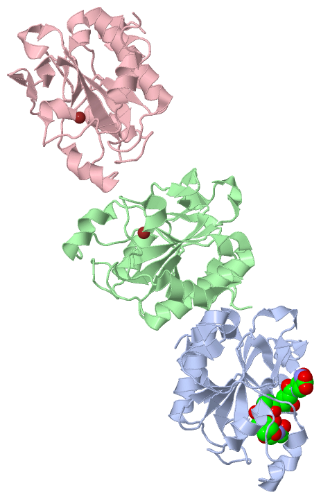
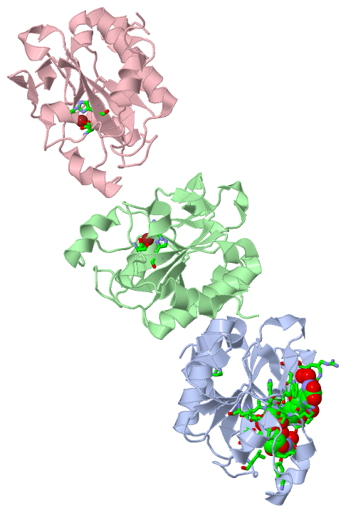
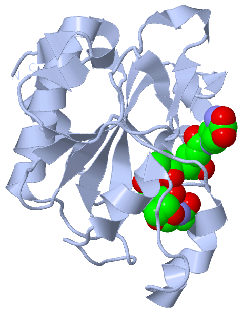
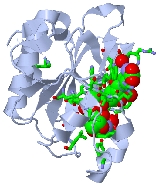
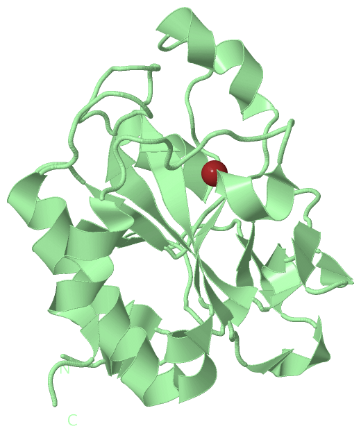
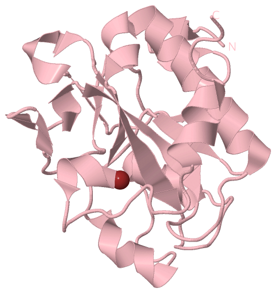
Asymmetric UnitChain A from PDB Type:PROTEIN Length:179 aligned with AMPD_CITFR | P82974 from UniProtKB/Swiss-Prot Length:187 Alignment length:179 10 20 30 40 50 60 70 80 90 100 110 120 130 140 150 160 170 AMPD_CITFR 1 MLLDEGWLAEARRVPSPHYDCRPDDENPSLLVVHNISLPPGEFGGPWIDALFTGTIDPNAHPYFAGIAHLRVSAHCLIRRDGEIVQYVPFDKRAWHAGVSSYQGRERCNDFSIGIELEGTDTLAYTDAQYQQLAAVTNALITRYPAIANNMTGHCNIAPERKTDPGPSFDWARFRALVT 179 SCOP domains d2y2ba_ A: AmpD protein SCOP domains CATH domains ----------------------------------------------------------------------------------------------------------------------------------------------------------------------------------- CATH domains Pfam domains ----------------------------------------------------------------------------------------------------------------------------------------------------------------------------------- Pfam domains SAPs(SNPs) ----------------------------------------------------------------------------------------------------------------------------------------------------------------------------------- SAPs(SNPs) PROSITE ----------------------------------------------------------------------------------------------------------------------------------------------------------------------------------- PROSITE Transcript ----------------------------------------------------------------------------------------------------------------------------------------------------------------------------------- Transcript 2y2b A 1 MLLDEGWLAEARRVPSPHYDCRPDDENPSLLVVHNISLPPGEFGGPWIDALFTGTIDPNAHPYFAGIAHLRVSAHCLIRRDGEIVQYVPFDKRAWHAGVSSYQGRERCNDFSIGIELEGTDTLAYTDAQYQQLAAVTNALITRYPAIANNMTGHCNIAPERKTDPGPSFDWARFRALVT 179 10 20 30 40 50 60 70 80 90 100 110 120 130 140 150 160 170 Chain B from PDB Type:PROTEIN Length:181 aligned with AMPD_CITFR | P82974 from UniProtKB/Swiss-Prot Length:187 Alignment length:181 10 20 30 40 50 60 70 80 90 100 110 120 130 140 150 160 170 180 AMPD_CITFR 1 MLLDEGWLAEARRVPSPHYDCRPDDENPSLLVVHNISLPPGEFGGPWIDALFTGTIDPNAHPYFAGIAHLRVSAHCLIRRDGEIVQYVPFDKRAWHAGVSSYQGRERCNDFSIGIELEGTDTLAYTDAQYQQLAAVTNALITRYPAIANNMTGHCNIAPERKTDPGPSFDWARFRALVTPS 181 SCOP domains d2y2bb_ B: AmpD protein SCOP domains CATH domains ------------------------------------------------------------------------------------------------------------------------------------------------------------------------------------- CATH domains Pfam domains ------------------------------------------------------------------------------------------------------------------------------------------------------------------------------------- Pfam domains SAPs(SNPs) ------------------------------------------------------------------------------------------------------------------------------------------------------------------------------------- SAPs(SNPs) PROSITE ------------------------------------------------------------------------------------------------------------------------------------------------------------------------------------- PROSITE Transcript ------------------------------------------------------------------------------------------------------------------------------------------------------------------------------------- Transcript 2y2b B 1 MLLDEGWLAEARRVPSPHYDCRPDDENPSLLVVHNISLPPGEFGGPWIDALFTGTIDPNAHPYFAGIAHLRVSAHCLIRRDGEIVQYVPFDKRAWHAGVSSYQGRERCNDFSIGIELEGTDTLAYTDAQYQQLAAVTNALITRYPAIANNMTGHCNIAPERKTDPGPSFDWARFRALVTPS 181 10 20 30 40 50 60 70 80 90 100 110 120 130 140 150 160 170 180 Chain C from PDB Type:PROTEIN Length:181 aligned with AMPD_CITFR | P82974 from UniProtKB/Swiss-Prot Length:187 Alignment length:181 10 20 30 40 50 60 70 80 90 100 110 120 130 140 150 160 170 180 AMPD_CITFR 1 MLLDEGWLAEARRVPSPHYDCRPDDENPSLLVVHNISLPPGEFGGPWIDALFTGTIDPNAHPYFAGIAHLRVSAHCLIRRDGEIVQYVPFDKRAWHAGVSSYQGRERCNDFSIGIELEGTDTLAYTDAQYQQLAAVTNALITRYPAIANNMTGHCNIAPERKTDPGPSFDWARFRALVTPS 181 SCOP domains d2y2bc_ C: AmpD protein SCOP domains CATH domains ------------------------------------------------------------------------------------------------------------------------------------------------------------------------------------- CATH domains Pfam domains ------------------------------------------------------------------------------------------------------------------------------------------------------------------------------------- Pfam domains SAPs(SNPs) ------------------------------------------------------------------------------------------------------------------------------------------------------------------------------------- SAPs(SNPs) PROSITE ------------------------------------------------------------------------------------------------------------------------------------------------------------------------------------- PROSITE Transcript ------------------------------------------------------------------------------------------------------------------------------------------------------------------------------------- Transcript 2y2b C 1 MLLDEGWLAEARRVPSPHYDCRPDDENPSLLVVHNISLPPGEFGGPWIDALFTGTIDPNAHPYFAGIAHLRVSAHCLIRRDGEIVQYVPFDKRAWHAGVSSYQGRERCNDFSIGIELEGTDTLAYTDAQYQQLAAVTNALITRYPAIANNMTGHCNIAPERKTDPGPSFDWARFRALVTPS 181 10 20 30 40 50 60 70 80 90 100 110 120 130 140 150 160 170 180
|
||||||||||||||||||||
SCOP Domains (1, 3)
Asymmetric Unit
 |
CATH Domains (0, 0)| (no "CATH Domain" information available for 2Y2B) |
Pfam Domains (0, 0)| (no "Pfam Domain" information available for 2Y2B) |
Gene Ontology (6, 6)|
Asymmetric Unit(hide GO term definitions) Chain A,B,C (AMPD_CITFR | P82974)
|
||||||||||||||||||||||||||||||||||||||||||||||||||||||
Interactive Views
|
||||||||||||||||||||||||||||||||||||||||||||||||||||||||||||||||||||||||||||||||||||||||||||||||||||||||||||||||||||||||||||||||||||||||||||||||||||||||||||||||||||||||||||||||||||||||||||||||||||||||||||||||||||||||||||||||
Still Images
|
||||||||||||||||
Databases
|
||||||||||||||||||||||||||||||||||||||||||||||||||||||||||||||||||||||||||||||||||||||||||||||||||||||||||||||||||||||||||||||||||||||||||||||||||||||||||||||||
Analysis Tools
|
|||||||||||||||||||||||||||||||||||||||||||||||||||||||||||||
Entries Sharing at Least One Protein Chain (UniProt ID)
Related Entries Specified in the PDB File
|
|