Show PDB file:   
         Plain Text   HTML   (compressed file size)
QuickSearch:   
by PDB,NDB,UniProt,PROSITE Code or Search Term(s)  
(-)NMR Structure - manually
(-)NMR Structure - model 1
(-)NMR Structure - all models
collapse expand < >
Image NMR Structure - manually
NMR Structure - manually  (Jmol Viewer)
Image NMR Structure - model 1
NMR Structure - model 1  (Jmol Viewer)
Image NMR Structure - all models
NMR Structure - all models  (Jmol Viewer)

(-) Description

Title :  SOLUTION STRUCTURE OF THE FKBP-DOMAIN OF LEGIONELLA PNEUMOPHILA MIP
 
Authors :  A. Ceymann, M. Horstmann, P. Ehses, K. Schweimer, M. Steinert, T. Kamph G. Fischer, J. Hacker, P. Rosch, C. Faber
Date :  25 Apr 07  (Deposition) - 12 Jun 07  (Release) - 14 Jun 17  (Revision)
Method :  SOLUTION NMR
Resolution :  NOT APPLICABLE
Chains :  NMR Structure  :  A  (10x)
NMR Structure *:  A  (1x)
Keywords :  Isomerase, Fkbp, Legionella Pneumophila, Ppiase, Legionnaires Disease (Keyword Search: [Gene Ontology, PubMed, Web (Google))
 
Reference :  M. Horstmann, P. Ehses, K. Schweimer, M. Steinert, T. Kamphausen, G. Fischer, J. Hacker, P. Rosch, C. Faber
Domain Motions Of The Mip Protein From Legionella Pneumophila
Biochemistry V. 45 12303 2006
PubMed-ID: 17014083  |  Reference-DOI: 10.1021/BI060818I
(for further references see the PDB file header)

(-) Compounds

Molecule 1 - MACROPHAGE INFECTIVITY POTENTIATOR
    ChainsA
    EC Number5.2.1.8
    FragmentFKBP DOMAIN, RESIDUES 97-233
    Organism ScientificLEGIONELLA PNEUMOPHILA
    Organism Taxid446
    Other DetailsNMR SOLUTION STRUCTURE OF THE MONOMERIC FKBP-DOMAIN OF MIP. IN THE REFERENCES THE N-TERMINAL 20 AMINO ACID SIGNAL SEQUENCE IS MISSING (ALL AMINO ACID NUMBERS THERE ARE MODIFIED WITH -20)
    Other Details - SourceSURFACE EXPOSED PROTEIN

 Structural Features

(-) Chains, Units

  1
NMR Structure (10x)A
NMR Structure * (1x)A

Summary Information (see also Sequences/Alignments below)

(-) Ligands, Modified Residues, Ions  (0, 0)

(no "Ligand,Modified Residues,Ions" information available for 2UZ5)

(-) Sites  (0, 0)

(no "Site" information available for 2UZ5)

(-) SS Bonds  (0, 0)

(no "SS Bond" information available for 2UZ5)

(-) Cis Peptide Bonds  (0, 0)

(no "Cis Peptide Bond" information available for 2UZ5)

 Sequence-Structure Mapping

(-) SAPs(SNPs)/Variants  (0, 0)

(no "SAP(SNP)/Variant" information available for 2UZ5)

(-) PROSITE Motifs  (1, 1)

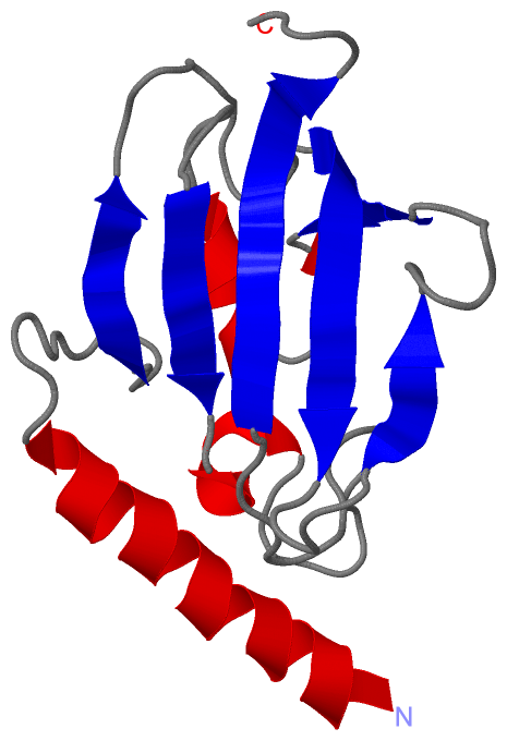
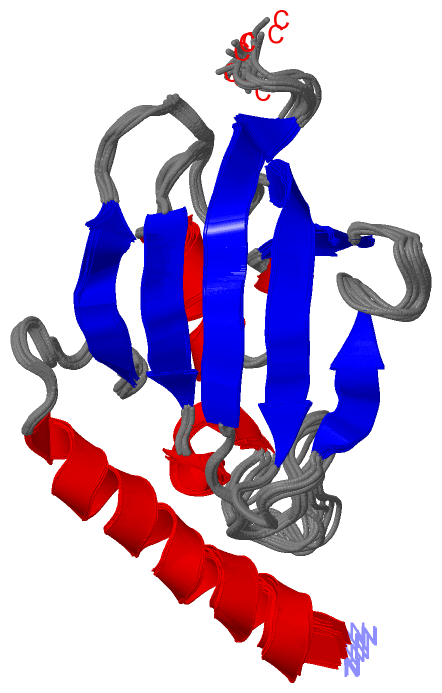
NMR Structure (1, 1)
 PROSITEUniProtKBPDB
No.IDACDescriptionIDLocationCountLocation
1FKBP_PPIASEPS50059 FKBP-type peptidyl-prolyl cis-trans isomerase domain profile.MIP_LEGPH144-230  1A:48-134
NMR Structure * (1, 1)
 PROSITEUniProtKBPDB
No.IDACDescriptionIDLocationCountLocation
1FKBP_PPIASEPS50059 FKBP-type peptidyl-prolyl cis-trans isomerase domain profile.MIP_LEGPH144-230  1A:48-134

(-) Exons   (0, 0)

(no "Exon" information available for 2UZ5)

(-) Sequences/Alignments

NMR Structure
   Reformat: Number of residues per line =  ('0' or empty: single-line sequence representation)
  Number of residues per labelling interval =   
  UniProt sequence: complete  aligned part    
   Show mapping: SCOP domains CATH domains Pfam domains Secondary structure (by author)
SAPs(SNPs) PROSITE motifs Exons
(details for a mapped element are shown in a popup box when the mouse pointer rests over it)
Chain A from PDB  Type:PROTEIN  Length:137
 aligned with MIP_LEGPH | Q5ZXE0 from UniProtKB/Swiss-Prot  Length:233

    Alignment length:137
                                   106       116       126       136       146       156       166       176       186       196       206       216       226       
            MIP_LEGPH    97 FNKKADENKVKGEAFLTENKNKPGVVVLPSGLQYKVINSGNGVKPGKSDTVTVEYTGRLIDGTVFDSTEKTGKPATFQVSQVIPGWTEALQLMPAGSTWEIYVPSGLAYGPRSVGGPIGPNETLIFKIHLISVKKSS 233
               SCOP domains d2uz5a_ A: automated matches                                                                                                              SCOP domains
               CATH domains ----------------------------------------------------------------------------------------------------------------------------------------- CATH domains
               Pfam domains FKBP_N-2uz5A02 A:1-36               ----FKBP_C-2uz5A01 A:41-131                                                                    ------ Pfam domains
         Sec.struct. author hhhhhhhhhhhhhhhhhhhh............eeeeee...........eeeeeeeee.....eeee.......eeee.hh..hhhhhhhh......eeeee.hhhhh................eeeeeeeee.... Sec.struct. author
                 SAPs(SNPs) ----------------------------------------------------------------------------------------------------------------------------------------- SAPs(SNPs)
                    PROSITE -----------------------------------------------FKBP_PPIASE  PDB: A:48-134 UniProt: 144-230                                            --- PROSITE
                 Transcript ----------------------------------------------------------------------------------------------------------------------------------------- Transcript
                 2uz5 A   1 FNKKADENKVKGEAFLTENKNKPGVVVLPSGLQYKVINSGNGVKPGKSDTVTVEYTGRLIDGTVFDSTEKTGKPATFQVSQVIPGWTEALQLMPAGSTWEIYVPSGLAYGPRSVGGPIGPNETLIFKIHLISVKKSS 137
                                    10        20        30        40        50        60        70        80        90       100       110       120       130       

Chain A from PDB  Type:PROTEIN  Length:137
 aligned with Q933L8_LEGPN | Q933L8 from UniProtKB/TrEMBL  Length:233

    Alignment length:137
                                   106       116       126       136       146       156       166       176       186       196       206       216       226       
         Q933L8_LEGPN    97 FNKKADENKVKGEAFLTENKNKPGVVVLPSGLQYKVINSGNGVKPGKSDTVTVEYTGRLIDGTVFDSTEKTGKPATFQVSQVIPGWTEALQLMPAGSTWEIYVPSGLAYGPRSVGGPIGPNETLIFKIHLISVKKSS 233
               SCOP domains d2uz5a_ A: automated matches                                                                                                              SCOP domains
               CATH domains ----------------------------------------------------------------------------------------------------------------------------------------- CATH domains
               Pfam domains FKBP_N-2uz5A02 A:1-36               ----FKBP_C-2uz5A01 A:41-131                                                                    ------ Pfam domains
         Sec.struct. author hhhhhhhhhhhhhhhhhhhh............eeeeee...........eeeeeeeee.....eeee.......eeee.hh..hhhhhhhh......eeeee.hhhhh................eeeeeeeee.... Sec.struct. author
                 SAPs(SNPs) ----------------------------------------------------------------------------------------------------------------------------------------- SAPs(SNPs)
                    PROSITE ----------------------------------------------------------------------------------------------------------------------------------------- PROSITE
                 Transcript ----------------------------------------------------------------------------------------------------------------------------------------- Transcript
                 2uz5 A   1 FNKKADENKVKGEAFLTENKNKPGVVVLPSGLQYKVINSGNGVKPGKSDTVTVEYTGRLIDGTVFDSTEKTGKPATFQVSQVIPGWTEALQLMPAGSTWEIYVPSGLAYGPRSVGGPIGPNETLIFKIHLISVKKSS 137
                                    10        20        30        40        50        60        70        80        90       100       110       120       130       

   Legend:   → Mismatch (orange background)
  - → Gap (green background, '-', border residues have a numbering label)
    → Modified Residue (blue background, lower-case, 'x' indicates undefined single-letter code, labelled with number + name)
  x → Chemical Group (purple background, 'x', labelled with number + name, e.g. ACE or NH2)
  extra numbering lines below/above indicate numbering irregularities and modified residue names etc., number ends below/above '|'

 Classification and Annotation

(-) SCOP Domains  (1, 1)

NMR Structure

(-) CATH Domains  (0, 0)

(no "CATH Domain" information available for 2UZ5)

(-) Pfam Domains  (2, 2)

NMR Structure

(-) Gene Ontology  (7, 12)

NMR Structure(hide GO term definitions)
Chain A   (MIP_LEGPH | Q5ZXE0)
molecular function
    GO:0016853    isomerase activity    Catalysis of the geometric or structural changes within one molecule. Isomerase is the systematic name for any enzyme of EC class 5.
    GO:0003755    peptidyl-prolyl cis-trans isomerase activity    Catalysis of the reaction: peptidyl-proline (omega=180) = peptidyl-proline (omega=0).
biological process
    GO:0009405    pathogenesis    The set of specific processes that generate the ability of an organism to induce an abnormal, generally detrimental state in another organism.
    GO:0006457    protein folding    The process of assisting in the covalent and noncovalent assembly of single chain polypeptides or multisubunit complexes into the correct tertiary structure.
    GO:0000413    protein peptidyl-prolyl isomerization    The modification of a protein by cis-trans isomerization of a proline residue.
cellular component
    GO:0009279    cell outer membrane    A lipid bilayer that forms the outermost membrane of the cell envelope; enriched in polysaccharide and protein; the outer leaflet of the membrane contains specific lipopolysaccharide structures.
    GO:0016020    membrane    A lipid bilayer along with all the proteins and protein complexes embedded in it an attached to it.

Chain A   (Q933L8_LEGPN | Q933L8)
molecular function
    GO:0016853    isomerase activity    Catalysis of the geometric or structural changes within one molecule. Isomerase is the systematic name for any enzyme of EC class 5.
    GO:0003755    peptidyl-prolyl cis-trans isomerase activity    Catalysis of the reaction: peptidyl-proline (omega=180) = peptidyl-proline (omega=0).
biological process
    GO:0006457    protein folding    The process of assisting in the covalent and noncovalent assembly of single chain polypeptides or multisubunit complexes into the correct tertiary structure.
    GO:0000413    protein peptidyl-prolyl isomerization    The modification of a protein by cis-trans isomerization of a proline residue.
cellular component
    GO:0016020    membrane    A lipid bilayer along with all the proteins and protein complexes embedded in it an attached to it.

 Visualization

(-) Interactive Views

NMR Structure
  Complete Structure
    Jena3D(integrated viewing of ligand, site, SAP, PROSITE, SCOP information)
    WebMol | AstexViewer[tm]@PDBe
(Java Applets, require no local installation except for Java; loading may be slow)
    STRAP
(Java WebStart application, automatic local installation, requires Java; full application with system access!)
    RasMol
(require local installation)
    Molscript (VRML)
(requires installation of a VRML viewer; select preferred view via VRML and generate a mono or stereo PDF format file)
 
  Ligands, Modified Residues, Ions
(no "Ligands, Modified Residues, Ions" information available for 2uz5)
 
  Sites
(no "Sites" information available for 2uz5)
 
  Cis Peptide Bonds
(no "Cis Peptide Bonds" information available for 2uz5)
 

(-) Still Images

Jmol
  protein: cartoon or spacefill or dots and stick; nucleic acid: cartoon and stick; ligands: spacefill; active site: stick
Molscript
  protein, nucleic acid: cartoon; ligands: spacefill; active site: ball and stick

 Databases and Analysis Tools

(-) Databases

Access by PDB/NDB ID
  2uz5
    Family and Domain InformationProDom | SYSTERS
    General Structural InformationGlycoscienceDB | MMDB | NDB | OCA | PDB | PDBe | PDBj | PDBsum | PDBWiki | PQS | PROTEOPEDIA
    Orientation in MembranesOPM
    Protein SurfaceSURFACE
    Secondary StructureDSSP (structure derived) | HSSP (homology derived)
    Structural GenomicsGeneCensus
    Structural NeighboursCE | VAST
    Structure ClassificationCATH | Dali | SCOP
    Validation and Original DataBMRB Data View | BMRB Restraints Grid | EDS | PROCHECK | RECOORD | WHAT_CHECK
 
Access by UniProt ID/Accession number
  MIP_LEGPH | Q5ZXE0
    Comparative Protein Structure ModelsModBase
    Genomic InformationEnsembl
    Protein-protein InteractionDIP
    Sequence, Family and Domain InformationInterPro | Pfam | SMART | UniProtKB/SwissProt
  Q933L8_LEGPN | Q933L8
    Comparative Protein Structure ModelsModBase
    Genomic InformationEnsembl
    Protein-protein InteractionDIP
    Sequence, Family and Domain InformationInterPro | Pfam | SMART | UniProtKB/SwissProt
 
Access by Enzyme Classificator   (EC Number)
  5.2.1.8
    General Enzyme InformationBRENDA | EC-PDB | Enzyme | IntEnz
    PathwayKEGG | MetaCyc
 
Access by Disease Identifier   (MIM ID)
  (no 'MIM ID' available)
    Disease InformationOMIM
 
Access by GenAge ID
  (no 'GenAge ID' available)
    Age Related InformationGenAge

(-) Analysis Tools

Access by PDB/NDB ID
    Domain InformationXDom
    Interatomic Contacts of Structural UnitsCSU
    Ligand-protein ContactsLPC
    Protein CavitiescastP
    Sequence and Secondary StructurePDBCartoon
    Structure AlignmentSTRAP(Java WebStart application, automatic local installation, requires Java; full application with system access!)
    Structure and Sequence BrowserSTING
 
Access by UniProt ID/Accession number
  MIP_LEGPH | Q5ZXE0
    Protein Disorder PredictionDisEMBL | FoldIndex | GLOBPLOT (for more information see DisProt)
  Q933L8_LEGPN | Q933L8
    Protein Disorder PredictionDisEMBL | FoldIndex | GLOBPLOT (for more information see DisProt)

 Related Entries

(-) Entries Sharing at Least One Protein Chain (UniProt ID)

UniProtKB/Swiss-Prot
        MIP_LEGPH | Q5ZXE01fd9 2vcd

(-) Related Entries Specified in the PDB File

(no "Related Entries Specified in the PDB File" available for 2UZ5)