Show PDB file:   
         Plain Text   HTML   (compressed file size)
QuickSearch:   
by PDB,NDB,UniProt,PROSITE Code or Search Term(s)  
(-)NMR Structure - model 1
(-)NMR Structure - all models
collapse expand < >
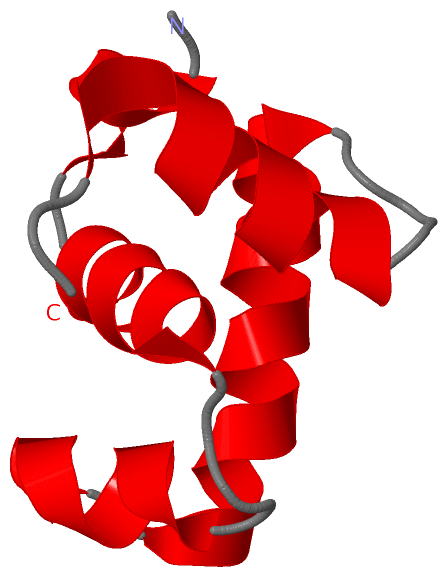
Image NMR Structure - model 1
NMR Structure - model 1  (Jmol Viewer)
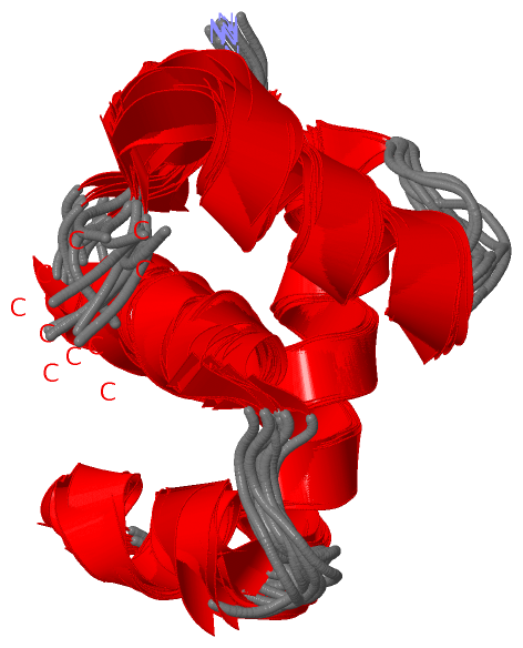
Image NMR Structure - all models
NMR Structure - all models  (Jmol Viewer)

(-) Description

Title :  NMR STRUCTURE OF THE S. AUREUS VRAR DNA BINDING DOMAIN
 
Authors :  L. W. Donaldson
Date :  09 Jan 08  (Deposition) - 22 Jan 08  (Release) - 24 Feb 09  (Revision)
Method :  SOLUTION NMR
Resolution :  NOT APPLICABLE
Chains :  NMR Structure  :  A  (10x)
Keywords :  Hth Luxr-Type Domain, Dna Binding Domain, Activator, Antibiotic Resistance, Cytoplasm, Dna-Binding, Phosphoprotein, Phosphorylation, Transcription, Transcription Regulation, Two-Component Regulatory System (Keyword Search: [Gene Ontology, PubMed, Web (Google))
 
Reference :  L. W. Donaldson
The Nmr Structure Of The Staphylococcus Aureus Response Regulator Vrar Dna Binding Domain Reveals A Dynamic Relationship Between It And Its Associated Receiver Domain
Biochemistry V. 47 3379 2008
PubMed-ID: 18293926  |  Reference-DOI: 10.1021/BI701844Q
(for further references see the PDB file header)

(-) Compounds

Molecule 1 - RESPONSE REGULATOR PROTEIN VRAR
    ChainsA
    EngineeredYES
    Expression SystemESCHERICHIA COLI BL21(DE3)
    Expression System Taxid469008
    Expression System VectorPET15B
    Expression System Vector TypeVECTOR
    FragmentHTH LUXR-TYPE DOMAIN, UNP RESIDUES 138-209
    GeneVRAR
    Organism ScientificSTAPHYLOCOCCUS AUREUS
    Organism Taxid1280
    Other DetailsCLONED AS NDEI/BAMHI FRAGMENT
    StrainMU50

 Structural Features

(-) Chains, Units

  
NMR Structure (10x)

Summary Information (see also Sequences/Alignments below)

(-) Ligands, Modified Residues, Ions  (0, 0)

(no "Ligand,Modified Residues,Ions" information available for 2RNJ)

(-) Sites  (0, 0)

(no "Site" information available for 2RNJ)

(-) SS Bonds  (0, 0)

(no "SS Bond" information available for 2RNJ)

(-) Cis Peptide Bonds  (0, 0)

(no "Cis Peptide Bond" information available for 2RNJ)

 Sequence-Structure Mapping

(-) SAPs(SNPs)/Variants  (0, 0)

(no "SAP(SNP)/Variant" information available for 2RNJ)

(-) PROSITE Motifs  (0, 0)

(no "PROSITE Motif" information available for 2RNJ)

(-) Exons   (0, 0)

(no "Exon" information available for 2RNJ)

(-) Sequences/Alignments

NMR Structure
   Reformat: Number of residues per line =  ('0' or empty: single-line sequence representation)
  Number of residues per labelling interval =   
  UniProt sequence: complete  aligned part    
   Show mapping: SCOP domains CATH domains Pfam domains Secondary structure (by author)
SAPs(SNPs) PROSITE motifs Exons
(details for a mapped element are shown in a popup box when the mouse pointer rests over it)
Chain A from PDB  Type:PROTEIN  Length:67
 aligned with VRAR_STAAM | Q7A2Q1 from UniProtKB/Swiss-Prot  Length:209

    Alignment length:67
                                   152       162       172       182       192       202       
           VRAR_STAAM   143 ELYEMLTEREMEILLLIAKGYSNQEIASASHITIKTVKTHVSNILSKLEVQDRTQAVIYAFQHNLIQ 209
               SCOP domains d2rnja_ A: automated matches                                        SCOP domains
               CATH domains 2rnjA00 A:143-209 'winged helix' repressor DNA binding domain       CATH domains
               Pfam domains --GerE-2rnjA01 A:145-202                                    ------- Pfam domains
         Sec.struct. author .hhhhh.hhhhhhhhhhhh....hhhhhhhh.hhhhhhhhhhhhhhhh....hhhhhhhhhhhh... Sec.struct. author
                 SAPs(SNPs) ------------------------------------------------------------------- SAPs(SNPs)
                    PROSITE ------------------------------------------------------------------- PROSITE
                 Transcript ------------------------------------------------------------------- Transcript
                 2rnj A 143 ELYEMLTEREMEILLLIAKGYSNQEIASASHITIKTVKTHVSNILSKLEVQDRTQAVIYAFQHNLIQ 209
                                   152       162       172       182       192       202       

   Legend:   → Mismatch (orange background)
  - → Gap (green background, '-', border residues have a numbering label)
    → Modified Residue (blue background, lower-case, 'x' indicates undefined single-letter code, labelled with number + name)
  x → Chemical Group (purple background, 'x', labelled with number + name, e.g. ACE or NH2)
  extra numbering lines below/above indicate numbering irregularities and modified residue names etc., number ends below/above '|'

 Classification and Annotation

(-) SCOP Domains  (1, 1)

NMR Structure

(-) CATH Domains  (1, 1)

NMR Structure

(-) Pfam Domains  (1, 1)

NMR Structure
(-)
Clan: HTH (544)

(-) Gene Ontology  (6, 6)

NMR Structure(hide GO term definitions)
Chain A   (VRAR_STAAM | Q7A2Q1)
molecular function
    GO:0003677    DNA binding    Any molecular function by which a gene product interacts selectively and non-covalently with DNA (deoxyribonucleic acid).
biological process
    GO:0000160    phosphorelay signal transduction system    A conserved series of molecular signals found in prokaryotes and eukaryotes; involves autophosphorylation of a histidine kinase and the transfer of the phosphate group to an aspartate that then acts as a phospho-donor to response regulator proteins.
    GO:0006355    regulation of transcription, DNA-templated    Any process that modulates the frequency, rate or extent of cellular DNA-templated transcription.
    GO:0046677    response to antibiotic    Any process that results in a change in state or activity of a cell or an organism (in terms of movement, secretion, enzyme production, gene expression, etc.) as a result of an antibiotic stimulus. An antibiotic is a chemical substance produced by a microorganism which has the capacity to inhibit the growth of or to kill other microorganisms.
    GO:0006351    transcription, DNA-templated    The cellular synthesis of RNA on a template of DNA.
cellular component
    GO:0005737    cytoplasm    All of the contents of a cell excluding the plasma membrane and nucleus, but including other subcellular structures.

 Visualization

(-) Interactive Views

NMR Structure
  Complete Structure
    Jena3D(integrated viewing of ligand, site, SAP, PROSITE, SCOP information)
    WebMol | AstexViewer[tm]@PDBe
(Java Applets, require no local installation except for Java; loading may be slow)
    STRAP
(Java WebStart application, automatic local installation, requires Java; full application with system access!)
    RasMol
(require local installation)
    Molscript (VRML)
(requires installation of a VRML viewer; select preferred view via VRML and generate a mono or stereo PDF format file)
 
  Ligands, Modified Residues, Ions
(no "Ligands, Modified Residues, Ions" information available for 2rnj)
 
  Sites
(no "Sites" information available for 2rnj)
 
  Cis Peptide Bonds
(no "Cis Peptide Bonds" information available for 2rnj)
 

(-) Still Images

Jmol
  protein: cartoon or spacefill or dots and stick; nucleic acid: cartoon and stick; ligands: spacefill; active site: stick
Molscript
  protein, nucleic acid: cartoon; ligands: spacefill; active site: ball and stick

 Databases and Analysis Tools

(-) Databases

Access by PDB/NDB ID
  2rnj
    Family and Domain InformationProDom | SYSTERS
    General Structural InformationGlycoscienceDB | MMDB | NDB | OCA | PDB | PDBe | PDBj | PDBsum | PDBWiki | PQS | PROTEOPEDIA
    Orientation in MembranesOPM
    Protein SurfaceSURFACE
    Secondary StructureDSSP (structure derived) | HSSP (homology derived)
    Structural GenomicsGeneCensus
    Structural NeighboursCE | VAST
    Structure ClassificationCATH | Dali | SCOP
    Validation and Original DataBMRB Data View | BMRB Restraints Grid | EDS | PROCHECK | RECOORD | WHAT_CHECK
 
Access by UniProt ID/Accession number
  VRAR_STAAM | Q7A2Q1
    Comparative Protein Structure ModelsModBase
    Genomic InformationEnsembl
    Protein-protein InteractionDIP
    Sequence, Family and Domain InformationInterPro | Pfam | SMART | UniProtKB/SwissProt
 
Access by Enzyme Classificator   (EC Number)
  (no 'Enzyme Classificator' available)
    General Enzyme InformationBRENDA | EC-PDB | Enzyme | IntEnz
    PathwayKEGG | MetaCyc
 
Access by Disease Identifier   (MIM ID)
  (no 'MIM ID' available)
    Disease InformationOMIM
 
Access by GenAge ID
  (no 'GenAge ID' available)
    Age Related InformationGenAge

(-) Analysis Tools

Access by PDB/NDB ID
    Domain InformationXDom
    Interatomic Contacts of Structural UnitsCSU
    Ligand-protein ContactsLPC
    Protein CavitiescastP
    Sequence and Secondary StructurePDBCartoon
    Structure AlignmentSTRAP(Java WebStart application, automatic local installation, requires Java; full application with system access!)
    Structure and Sequence BrowserSTING
 
Access by UniProt ID/Accession number
  VRAR_STAAM | Q7A2Q1
    Protein Disorder PredictionDisEMBL | FoldIndex | GLOBPLOT (for more information see DisProt)

 Related Entries

(-) Entries Sharing at Least One Protein Chain (UniProt ID)

UniProtKB/Swiss-Prot
        VRAR_STAAM | Q7A2Q14gvp 4if4

(-) Related Entries Specified in the PDB File

(no "Related Entries Specified in the PDB File" available for 2RNJ)