|
|
|
|
 Description
|
|
Compounds
|
||||||||||||||||||||||||
Chains, Units
Summary Information (see also Sequences/Alignments below) |
Ligands, Modified Residues, Ions (6, 7)
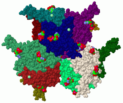
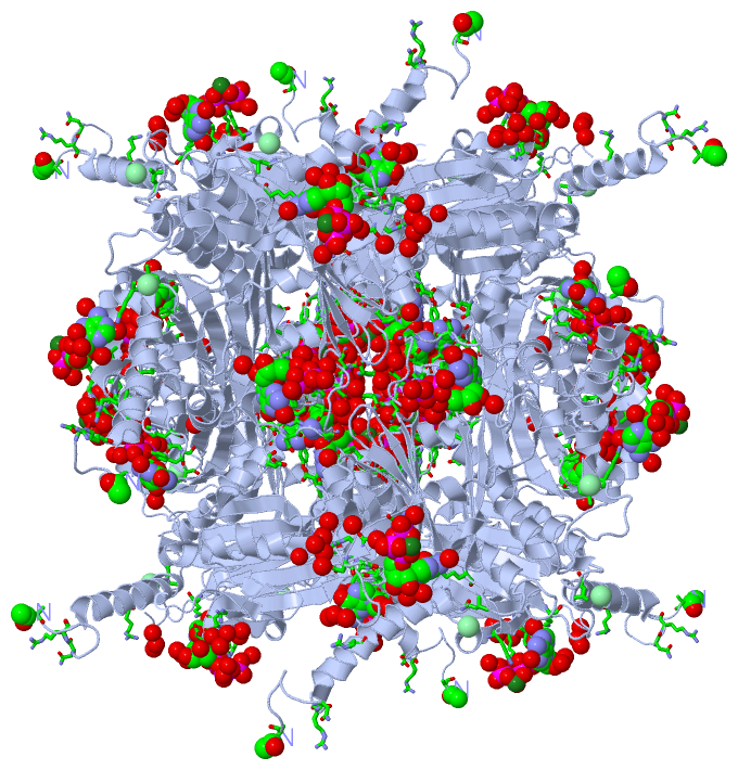
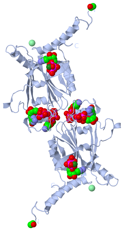
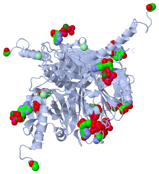
Asymmetric Unit (6, 7)
|
Sites (6, 6)
Asymmetric Unit (6, 6)
|
SS Bonds (0, 0)| (no "SS Bond" information available for 2OH5) |
Cis Peptide Bonds (0, 0)| (no "Cis Peptide Bond" information available for 2OH5) |
SAPs(SNPs)/Variants (0, 0)| (no "SAP(SNP)/Variant" information available for 2OH5) |
PROSITE Motifs (0, 0)| (no "PROSITE Motif" information available for 2OH5) |
Exons (0, 0)| (no "Exon" information available for 2OH5) |
Sequences/Alignments
Asymmetric UnitChain A from PDB Type:PROTEIN Length:248 aligned with O10693_CPVBM | O10693 from UniProtKB/TrEMBL Length:248 Alignment length:248 10 20 30 40 50 60 70 80 90 100 110 120 130 140 150 160 170 180 190 200 210 220 230 240 O10693_CPVBM 1 MADVAGTSNRDFRGREQRLFNSEQYNYNNSLNGEVSVWVYAYYSDGSVLVINKNSQYKVGISETFKALKEYRKGQHNDSYDEYEVNQSIYYPNGGDARKFHSNAKPRAIQIIFSPSVNVRTIKMAKGNAVSVPDEYLQRSHPWEATGIKYRKIKRDGEIVGYSHYFELPHEYNSISLAVSGVHKNPSSYNVGSAHNVMDVFQSCDLALRFCNRYWAELELVNHYISPNAYPYLDINNHSYGVALSNRQ 248 SCOP domains -------------------------------------------------------------------------------------------------------------------------------------------------------------------------------------------------------------------------------------------------------- SCOP domains CATH domains -------------------------------------------------------------------------------------------------------------------------------------------------------------------------------------------------------------------------------------------------------- CATH domains Pfam domains -Cypo_polyhedrin-2oh5A01 A:2-248 Pfam domains SAPs(SNPs) -------------------------------------------------------------------------------------------------------------------------------------------------------------------------------------------------------------------------------------------------------- SAPs(SNPs) PROSITE -------------------------------------------------------------------------------------------------------------------------------------------------------------------------------------------------------------------------------------------------------- PROSITE Transcript -------------------------------------------------------------------------------------------------------------------------------------------------------------------------------------------------------------------------------------------------------- Transcript 2oh5 A 1 xADVAGTSNRDFRGREQRLFNSEQYNYNNSLNGEVSVWVYAYYSDGSVLVINKNSQYKVGISETFKALKEYRKGQHNDSYDEYEVNQSIYYPNGGDARKFHSNAKPRAIQIIFSPSVNVRTIKMAKGNAVSVPDEYLQRSHPWEATGIKYRKIKRDGEIVGYSHYFELPHEYNSISLAVSGVHKNPSSYNVGSAHNVMDVFQSCDLALRFCNRYWAELELVNHYISPNAYPYLDINNHSYGVALSNRQ 248 | 10 20 30 40 50 60 70 80 90 100 110 120 130 140 150 160 170 180 190 200 210 220 230 240 | 1-ACE
|
||||||||||||||||||||
SCOP Domains (0, 0)| (no "SCOP Domain" information available for 2OH5) |
CATH Domains (0, 0)| (no "CATH Domain" information available for 2OH5) |
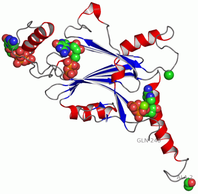
Pfam Domains (1, 1)
Asymmetric Unit
|
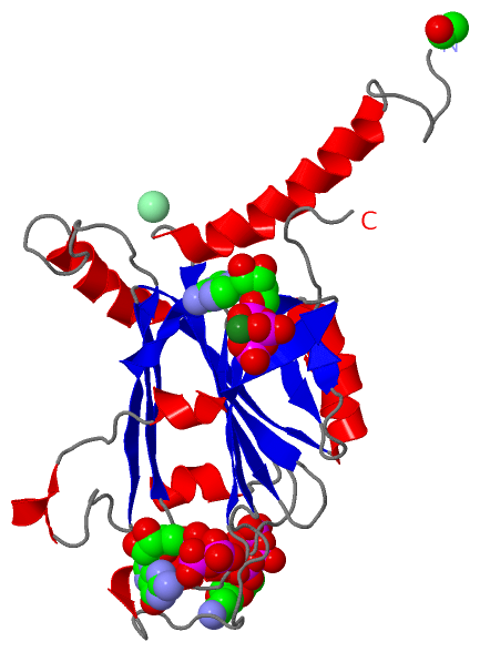
Gene Ontology (3, 3)|
Asymmetric Unit(hide GO term definitions) Chain A (O10693_CPVBM | O10693)
|
||||||||||||||||||||||||
Interactive Views
|
||||||||||||||||||||||||||||||||||||||||||||||||||||||||||||||||||||||||||||||||||||||||||||||||||||||||||||||||||||||||||||||||||||||||||||||||||||||||||||||||||||||||||||||||||||||||||||||||||||||||||||||||||||||||
Still Images
|
||||||||||||||||
Databases
|
||||||||||||||||||||||||||||||||||||||||||||||||||||||||||||||||||||||||||||||||||||||||||||||||||||||||||||||||||||||||||||||||||||||||||||||||||||||||||||||||
Analysis Tools
|
|||||||||||||||||||||||||||||||||||||||||||||||||||||||||||||
Entries Sharing at Least One Protein Chain (UniProt ID)
Related Entries Specified in the PDB File
|
|