|
|
|
|
 Description
|
|
Compounds
|
||||||||||||||||||||||||||||||||||||||||||||||||
Chains, Units
Summary Information (see also Sequences/Alignments below) |
Ligands, Modified Residues, Ions (4, 5)| Asymmetric Unit (4, 5) Biological Unit 1 (3, 12) |
Sites (5, 5)
Asymmetric Unit (5, 5)
|
SS Bonds (0, 0)| (no "SS Bond" information available for 2NT8) |
Cis Peptide Bonds (0, 0)| (no "Cis Peptide Bond" information available for 2NT8) |
SAPs(SNPs)/Variants (0, 0)| (no "SAP(SNP)/Variant" information available for 2NT8) |
PROSITE Motifs (0, 0)| (no "PROSITE Motif" information available for 2NT8) |
Exons (0, 0)| (no "Exon" information available for 2NT8) |
Sequences/Alignments
Asymmetric UnitChain A from PDB Type:PROTEIN Length:181 aligned with Q50EJ2_LACRE | Q50EJ2 from UniProtKB/TrEMBL Length:188 Alignment length:181 11 21 31 41 51 61 71 81 91 101 111 121 131 141 151 161 171 181 Q50EJ2_LACRE 2 KIYTKNGDKGQTRIIGKQILYKNDPRVAAYGEVDELNSWVGYTKSLINSHTQVLSNELEEIQQLLFDCGHDLATPADDERHSFKFKQEQPTVWLEEKIDNYTQVVPAVKKFILPGGTQLASALHVARTITRRAERQIVQLMREEQINQDVLIFINRLSDYFFAAARYANYLEQQPDMLYRN 182 SCOP domains d2nt8a_ A: automated matches SCOP domains CATH domains ----------2nt8A01 A:12-182 [code=1.20.1200.10, no name defined] CATH domains Pfam domains -Cob_adeno_trans-2nt8A01 A:3-170 ------------ Pfam domains SAPs(SNPs) ------------------------------------------------------------------------------------------------------------------------------------------------------------------------------------- SAPs(SNPs) PROSITE ------------------------------------------------------------------------------------------------------------------------------------------------------------------------------------- PROSITE Transcript ------------------------------------------------------------------------------------------------------------------------------------------------------------------------------------- Transcript 2nt8 A 2 KIYTKNGDKGQTRIIGKQILYKNDPRVAAYGEVDELNSWVGYTKSLINSHTQVLSNELEEIQQLLFDCGHDLATPADDERHSFKFKQEQPTVWLEEKIDNYTQVVPAVKKFILPGGTQLASALHVARTITRRAERQIVQLMREEQINQDVLIFINRLSDYFFAAARYANYLEQQPDMLYRN 182 11 21 31 41 51 61 71 81 91 101 111 121 131 141 151 161 171 181
|
||||||||||||||||||||
SCOP Domains (1, 1)
Asymmetric Unit
 |
CATH Domains (1, 1)
Asymmetric Unit
|
Pfam Domains (1, 1)
Asymmetric Unit
|
Gene Ontology (5, 5)|
Asymmetric Unit(hide GO term definitions) Chain A (Q50EJ2_LACRE | Q50EJ2)
|
||||||||||||||||||||||||||||||||||||
Interactive Views
|
|||||||||||||||||||||||||||||||||||||||||||||||||||||||||||||||||||||||||||||||||||||||||||||||||||||||||||||||||||||||||||||||||||||||||||||||||||||||||||||||||||||||||||||||||||||||||
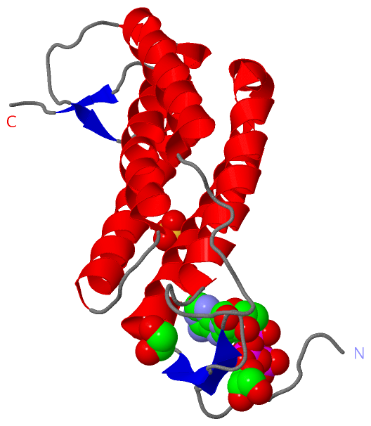
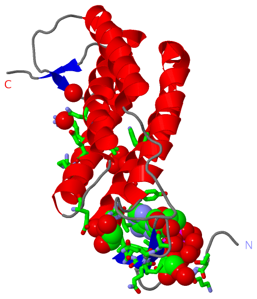
Still Images
|
||||||||||||||||
Databases
|
||||||||||||||||||||||||||||||||||||||||||||||||||||||||||||||||||||||||||||||||||||||||||||||||||||||||||||||||||||||||||||||||||||||||||||||||||||||||||||||||
Analysis Tools
|
|||||||||||||||||||||||||||||||||||||||||||||||||||||||||||||
Entries Sharing at Least One Protein Chain (UniProt ID)
Related Entries Specified in the PDB File
|
|