|
|
|
|
 Description
|
|
Compounds
|
||||||||||||||||||||||||||||||||||||||||||||||||||||||||
Chains, Units
Summary Information (see also Sequences/Alignments below) |
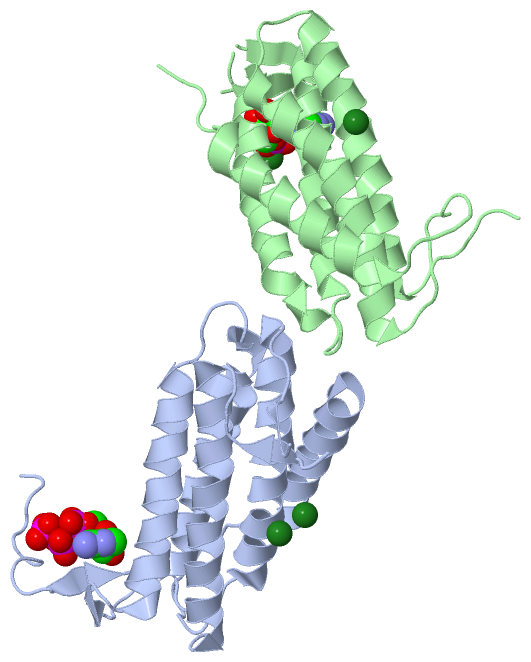
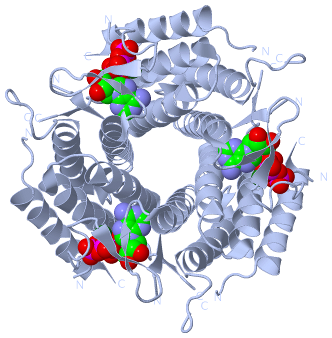
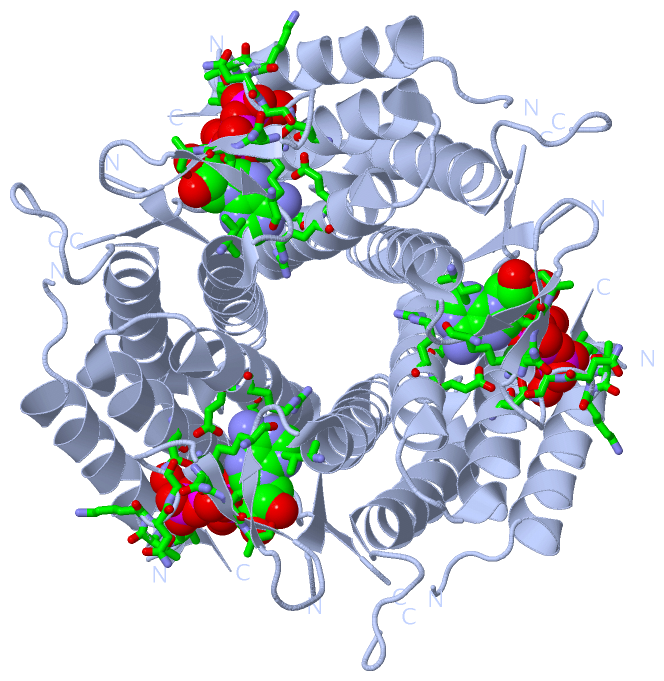
Ligands, Modified Residues, Ions (2, 6)| Asymmetric Unit (2, 6) Biological Unit 1 (1, 3) Biological Unit 2 (1, 3) |
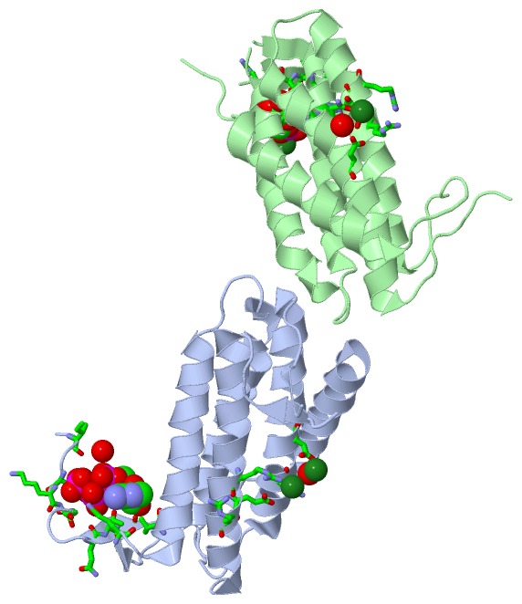
Sites (6, 6)
Asymmetric Unit (6, 6)
|
SS Bonds (0, 0)| (no "SS Bond" information available for 2R6X) |
Cis Peptide Bonds (1, 1)
Asymmetric Unit
|
||||||||
SAPs(SNPs)/Variants (0, 0)| (no "SAP(SNP)/Variant" information available for 2R6X) |
PROSITE Motifs (0, 0)| (no "PROSITE Motif" information available for 2R6X) |
Exons (0, 0)| (no "Exon" information available for 2R6X) |
Sequences/Alignments
Asymmetric UnitChain A from PDB Type:PROTEIN Length:176 aligned with Q50EJ2_LACRE | Q50EJ2 from UniProtKB/TrEMBL Length:188 Alignment length:180 11 21 31 41 51 61 71 81 91 101 111 121 131 141 151 161 171 181 Q50EJ2_LACRE 2 KIYTKNGDKGQTRIIGKQILYKNDPRVAAYGEVDELNSWVGYTKSLINSHTQVLSNELEEIQQLLFDCGHDLATPADDERHSFKFKQEQPTVWLEEKIDNYTQVVPAVKKFILPGGTQLASALHVARTITRRAERQIVQLMREEQINQDVLIFINRLSDYFFAAARYANYLEQQPDMLYR 181 SCOP domains d2r6xa_ A: automated matches SCOP domains CATH domains ----------2r6xA01 A:12-181 [code=1.20.1200.10, no name defined] CATH domains Pfam domains ------------------------------------------------------------------------------------------------------------------------------------------------------------------------------------ Pfam domains SAPs(SNPs) ------------------------------------------------------------------------------------------------------------------------------------------------------------------------------------ SAPs(SNPs) PROSITE ------------------------------------------------------------------------------------------------------------------------------------------------------------------------------------ PROSITE Transcript ------------------------------------------------------------------------------------------------------------------------------------------------------------------------------------ Transcript 2r6x A 2 KIYTKNGDKGQTRIIGKQILYKNDPRVAAYGEVNELNSWVGYTKSLINSHTQVLSNELEEIQQLLFDCGHDLATPADDERH-FKFKQEQPTVWLEEKIDNYTQVVP---KFILPGGTQLASALHVARTITRRAERQIVQLMREEQINQDVLIFINRLSDYFFAAARYANYLEQQPDMLYR 181 11 21 31 41 51 61 71 81| | 91 101 | 111 121 131 141 151 161 171 181 82 | 107 111 84 Chain B from PDB Type:PROTEIN Length:173 aligned with Q50EJ2_LACRE | Q50EJ2 from UniProtKB/TrEMBL Length:188 Alignment length:179 12 22 32 42 52 62 72 82 92 102 112 122 132 142 152 162 172 Q50EJ2_LACRE 3 IYTKNGDKGQTRIIGKQILYKNDPRVAAYGEVDELNSWVGYTKSLINSHTQVLSNELEEIQQLLFDCGHDLATPADDERHSFKFKQEQPTVWLEEKIDNYTQVVPAVKKFILPGGTQLASALHVARTITRRAERQIVQLMREEQINQDVLIFINRLSDYFFAAARYANYLEQQPDMLYR 181 SCOP domains d2r6xb_ B: aut omated matches SCOP domains CATH domains ---------2r6xB 01 B:12-181 [code=1.20.1200.10, no name defined] CATH domains Pfam domains (1) Cob_adeno_tran s-2r6xB01 B:3-170 ----------- Pfam domains (1) Pfam domains (2) Cob_adeno_tran s-2r6xB02 B:3-170 ----------- Pfam domains (2) SAPs(SNPs) ----------------------------------------------------------------------------------------------------------------------------------------------------------------------------------- SAPs(SNPs) PROSITE ----------------------------------------------------------------------------------------------------------------------------------------------------------------------------------- PROSITE Transcript ----------------------------------------------------------------------------------------------------------------------------------------------------------------------------------- Transcript 2r6x B 3 IYTKNGDKGQTRII--QILYKNDPRVAAYGEVNELNSWVGYTKSLINSHTQVLSNELEEIQQLLFDCGHDLATP----RHSFKFKQEQPTVWLEEKIDNYTQVVPAVKKFILPGGTQLASALHVARTITRRAERQIVQLMREEQINQDVLIFINRLSDYFFAAARYANYLEQQPDMLYR 181 12 | | 22 32 42 52 62 72 | 82 92 102 112 122 132 142 152 162 172 16 19 76 81
|
||||||||||||||||||||
SCOP Domains (1, 2)
Asymmetric Unit
 |
CATH Domains (1, 2)
Asymmetric Unit
|
Pfam Domains (1, 2)
Asymmetric Unit
|
Gene Ontology (5, 5)|
Asymmetric Unit(hide GO term definitions) Chain A,B (Q50EJ2_LACRE | Q50EJ2)
|
||||||||||||||||||||||||||||||||||||
Interactive Views
|
||||||||||||||||||||||||||||||||||||||||||||||||||||||||||||||||||||||||||||||||||||||||||||||||||||||||||||||||||||||||||||||||||||||||||||||||||||||||||||||||||||||||||||||||||||||||
Still Images
|
||||||||||||||||
Databases
|
||||||||||||||||||||||||||||||||||||||||||||||||||||||||||||||||||||||||||||||||||||||||||||||||||||||||||||||||||||||||||||||||||||||||||||||||||||||||||||||||
Analysis Tools
|
|||||||||||||||||||||||||||||||||||||||||||||||||||||||||||||
Entries Sharing at Least One Protein Chain (UniProt ID)
Related Entries Specified in the PDB File
|
|