|
|
|
|
 Description
|
|
Compounds
|
||||||||||||||||||||||||||||||||||||||||||||
Chains, Units
Summary Information (see also Sequences/Alignments below) |
Ligands, Modified Residues, Ions (1, 1)
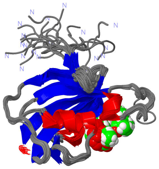
NMR Structure (1, 1)
|
Sites (1, 1)
NMR Structure (1, 1)
|
SS Bonds (0, 0)| (no "SS Bond" information available for 2KO7) |
Cis Peptide Bonds (0, 0)| (no "Cis Peptide Bond" information available for 2KO7) |
SAPs(SNPs)/Variants (0, 0)| (no "SAP(SNP)/Variant" information available for 2KO7) |
PROSITE Motifs (0, 0)| (no "PROSITE Motif" information available for 2KO7) |
Exons (0, 0)| (no "Exon" information available for 2KO7) |
Sequences/Alignments
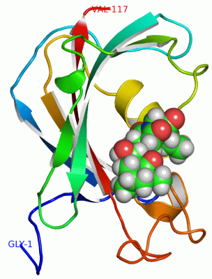
NMR StructureChain A from PDB Type:PROTEIN Length:117 aligned with Q63J95_BURPS | Q63J95 from UniProtKB/TrEMBL Length:113 Alignment length:117 1 | 6 16 26 36 46 56 66 76 86 96 106 Q63J95_BURPS - ----MTVVTTESGLKYEDLTEGSGAEARAGQTVSVHYTGWLTDGQKFDSSKDRNDPFAFVLGGGMVIKGWDEGVQGMKVGGVRRLTIPPQLGYGARGAGGVIPPNATLVFEVELLDV 113 SCOP domains d2ko7a_ A: automated matches SCOP domains CATH domains --------------------------------------------------------------------------------------------------------------------- CATH domains Pfam domains -----------------------FKBP_C-2ko7A01 A:24-115 -- Pfam domains SAPs(SNPs) --------------------------------------------------------------------------------------------------------------------- SAPs(SNPs) PROSITE --------------------------------------------------------------------------------------------------------------------- PROSITE Transcript --------------------------------------------------------------------------------------------------------------------- Transcript 2ko7 A 1 GPGSMTVVTTESGLKYEDLTEGSGAEARAGQTVSVHYTGWLTDGQKFDSSKDRNDPFAFVLGGGMVIKGWDEGVQGMKVGGVRRLTIPPQLGYGARGAGGVIPPNATLVFEVELLDV 117 10 20 30 40 50 60 70 80 90 100 110
|
||||||||||||||||||||
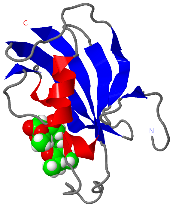
SCOP Domains (1, 1)
NMR Structure
 |
CATH Domains (0, 0)| (no "CATH Domain" information available for 2KO7) |
Pfam Domains (1, 1)
NMR Structure
|
Gene Ontology (4, 4)|
NMR Structure(hide GO term definitions) Chain A (Q63J95_BURPS | Q63J95)
|
||||||||||||||||||||||||||||||||||||
Interactive Views
|
||||||||||||||||||||||||||||||||||||||||||||||||||||||||||||||||||||||||||||||||||||||||||||||||||||||||||||||||||||||
Still Images
|
||||||||||||||||
Databases
|
||||||||||||||||||||||||||||||||||||||||||||||||||||||||||||||||||||||||||||||||||||||||||||||||||||||||||||||||||||||||||||||||||||||||||||||||||||||||||||||||
Analysis Tools
|
|||||||||||||||||||||||||||||||||||||||||||||||||||||||||||||
Entries Sharing at Least One Protein Chain (UniProt ID)
Related Entries Specified in the PDB File
|
|