Show PDB file:   
         Plain Text   HTML   (compressed file size)
QuickSearch:   
by PDB,NDB,UniProt,PROSITE Code or Search Term(s)  
(-)NMR Structure - model 1
(-)NMR Structure - all models
collapse expand < >
Image NMR Structure - model 1
NMR Structure - model 1  (Jmol Viewer)
Image NMR Structure - all models
NMR Structure - all models  (Jmol Viewer)

(-) Description

Title :  SOLUTION STRUCTURE OF THE N-TERMINAL DOMAIN OF PH1500
 
Authors :  I. Varnay, V. Truffault, M. Coles
Date :  11 Sep 07  (Deposition) - 25 Sep 07  (Release) - 24 Feb 09  (Revision)
Method :  SOLUTION NMR
Resolution :  NOT APPLICABLE
Chains :  NMR Structure  :  A  (22x)
Keywords :  Aaa Atpase Nc-Domain-Like, Unknown Function (Keyword Search: [Gene Ontology, PubMed, Web (Google))
 
Reference :  I. Varnay, S. Djuranovic, V. Truffault, A. Lupas, H. Kessler, M. Coles
The Solution Structure Of Ph1500-N
To Be Published
PubMed: search
(for further references see the PDB file header)

(-) Compounds

Molecule 1 - PUTATIVE UNCHARACTERIZED PROTEIN PH1500
    ChainsA
    EngineeredYES
    Expression SystemESCHERICHIA COLI
    Expression System Taxid562
    Expression System VectorPET30B
    Expression System Vector TypePLASMID
    GenePH1500
    Organism ScientificPYROCOCCUS HORIKOSHII
    Organism Taxid53953
    Other DetailsCIS-P66, CIS-P68

 Structural Features

(-) Chains, Units

  
NMR Structure (22x)

Summary Information (see also Sequences/Alignments below)

(-) Ligands, Modified Residues, Ions  (0, 0)

(no "Ligand,Modified Residues,Ions" information available for 2JV2)

(-) Sites  (0, 0)

(no "Site" information available for 2JV2)

(-) SS Bonds  (0, 0)

(no "SS Bond" information available for 2JV2)

(-) Cis Peptide Bonds  (2, 44)
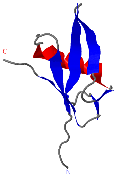
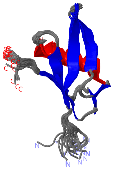
NMR Structure
No.ModelResidues
11, 2, 3, 4, 5, 6, 7, 8, 9, 10, 11, 12, 13, 14, 15, 16, 17, 18, 19, 20, 21, 22Tyr A:65 -Pro A:66
21, 2, 3, 4, 5, 6, 7, 8, 9, 10, 11, 12, 13, 14, 15, 16, 17, 18, 19, 20, 21, 22Ser A:67 -Pro A:68

 Sequence-Structure Mapping

(-) SAPs(SNPs)/Variants  (0, 0)

(no "SAP(SNP)/Variant" information available for 2JV2)

(-) PROSITE Motifs  (0, 0)

(no "PROSITE Motif" information available for 2JV2)

(-) Exons   (0, 0)

(no "Exon" information available for 2JV2)

(-) Sequences/Alignments

NMR Structure
   Reformat: Number of residues per line =  ('0' or empty: single-line sequence representation)
  Number of residues per labelling interval =   
  UniProt sequence: complete  aligned part    
   Show mapping: SCOP domains CATH domains Pfam domains Secondary structure (by author)
SAPs(SNPs) PROSITE motifs Exons
(details for a mapped element are shown in a popup box when the mouse pointer rests over it)
Chain A from PDB  Type:PROTEIN  Length:76
 aligned with O59169_PYRHO | O59169 from UniProtKB/TrEMBL  Length:148

    Alignment length:76
                                    11        21        31        41        51        61        71      
          O59169_PYRHO    2 EGVIMSELKLKPLPKVELPPDFVDVIRIKLQGKTVRTGDVIGISILGKEVKFKVVQAYPSPLRVEDRTKITLVTHP 77
               SCOP domains ---------------------------------------------------------------------------- SCOP domains
               CATH domains ---------------------------------------------------------------------------- CATH domains
               Pfam domains ----------------CDC48_2-2jv2A01 A:24-83                                      Pfam domains
         Sec.struct. author ......eeeeee......hhhhhhhhhhhhh..ee....eeeeee..eeeeeeeeeee...ee.....eeee.... Sec.struct. author
                 SAPs(SNPs) ---------------------------------------------------------------------------- SAPs(SNPs)
                    PROSITE ---------------------------------------------------------------------------- PROSITE
                 Transcript ---------------------------------------------------------------------------- Transcript
                  2jv2 A  8 EGVIMSELKLKPLPKVELPPDFVDVIRIKLQGKTVRTGDVIGISILGKEVKFKVVQAYPSPLRVEDRTKITLVTHP 83
                                    17        27        37        47        57        67        77      

   Legend:   → Mismatch (orange background)
  - → Gap (green background, '-', border residues have a numbering label)
    → Modified Residue (blue background, lower-case, 'x' indicates undefined single-letter code, labelled with number + name)
  x → Chemical Group (purple background, 'x', labelled with number + name, e.g. ACE or NH2)
  extra numbering lines below/above indicate numbering irregularities and modified residue names etc., number ends below/above '|'

 Classification and Annotation

(-) SCOP Domains  (0, 0)

(no "SCOP Domain" information available for 2JV2)

(-) CATH Domains  (0, 0)

(no "CATH Domain" information available for 2JV2)

(-) Pfam Domains  (1, 1)

NMR Structure

(-) Gene Ontology  (0, 0)

NMR Structure(hide GO term definitions)
    (no "Gene Ontology" information available for 2JV2)

 Visualization

(-) Interactive Views

NMR Structure
  Complete Structure
    Jena3D(integrated viewing of ligand, site, SAP, PROSITE, SCOP information)
    WebMol | AstexViewer[tm]@PDBe
(Java Applets, require no local installation except for Java; loading may be slow)
    STRAP
(Java WebStart application, automatic local installation, requires Java; full application with system access!)
    RasMol
(require local installation)
    Molscript (VRML)
(requires installation of a VRML viewer; select preferred view via VRML and generate a mono or stereo PDF format file)
 
  Ligands, Modified Residues, Ions
(no "Ligands, Modified Residues, Ions" information available for 2jv2)
 
  Sites
(no "Sites" information available for 2jv2)
 
  Cis Peptide Bonds
    Ser A:67 - Pro A:68   [ RasMol ]  
    Tyr A:65 - Pro A:66   [ RasMol ]  
 

(-) Still Images

Jmol
  protein: cartoon or spacefill or dots and stick; nucleic acid: cartoon and stick; ligands: spacefill; active site: stick
Molscript
  protein, nucleic acid: cartoon; ligands: spacefill; active site: ball and stick

 Databases and Analysis Tools

(-) Databases

Access by PDB/NDB ID
  2jv2
    Family and Domain InformationProDom | SYSTERS
    General Structural InformationGlycoscienceDB | MMDB | NDB | OCA | PDB | PDBe | PDBj | PDBsum | PDBWiki | PQS | PROTEOPEDIA
    Orientation in MembranesOPM
    Protein SurfaceSURFACE
    Secondary StructureDSSP (structure derived) | HSSP (homology derived)
    Structural GenomicsGeneCensus
    Structural NeighboursCE | VAST
    Structure ClassificationCATH | Dali | SCOP
    Validation and Original DataBMRB Data View | BMRB Restraints Grid | EDS | PROCHECK | RECOORD | WHAT_CHECK
 
Access by UniProt ID/Accession number
  O59169_PYRHO | O59169
    Comparative Protein Structure ModelsModBase
    Genomic InformationEnsembl
    Protein-protein InteractionDIP
    Sequence, Family and Domain InformationInterPro | Pfam | SMART | UniProtKB/TrEMBL
 
Access by Enzyme Classificator   (EC Number)
  (no 'Enzyme Classificator' available)
    General Enzyme InformationBRENDA | EC-PDB | Enzyme | IntEnz
    PathwayKEGG | MetaCyc
 
Access by Disease Identifier   (MIM ID)
  (no 'MIM ID' available)
    Disease InformationOMIM
 
Access by GenAge ID
  (no 'GenAge ID' available)
    Age Related InformationGenAge

(-) Analysis Tools

Access by PDB/NDB ID
    Domain InformationXDom
    Interatomic Contacts of Structural UnitsCSU
    Ligand-protein ContactsLPC
    Protein CavitiescastP
    Sequence and Secondary StructurePDBCartoon
    Structure AlignmentSTRAP(Java WebStart application, automatic local installation, requires Java; full application with system access!)
    Structure and Sequence BrowserSTING
 
Access by UniProt ID/Accession number
  O59169_PYRHO | O59169
    Protein Disorder PredictionDisEMBL | FoldIndex | GLOBPLOT (for more information see DisProt)

 Related Entries

(-) Entries Sharing at Least One Protein Chain (UniProt ID)

UniProtKB/TrEMBL
        O59169_PYRHO | O591692m3x 4uzr

(-) Related Entries Specified in the PDB File

(no "Related Entries Specified in the PDB File" available for 2JV2)