Show PDB file:   
         Plain Text   HTML   (compressed file size)
QuickSearch:   
by PDB,NDB,UniProt,PROSITE Code or Search Term(s)  
(-)NMR Structure - manually
(-)NMR Structure - model 1
(-)NMR Structure - model 1, sites
(-)NMR Structure - all models
collapse expand < >
Image NMR Structure - manually
NMR Structure - manually  (Jmol Viewer)
Image NMR Structure - model 1
NMR Structure - model 1  (Jmol Viewer)
Image NMR Structure - model 1, sites
NMR Structure - model 1, sites  (Jmol Viewer)
Image NMR Structure - all models
NMR Structure - all models  (Jmol Viewer)

(-) Description

Title :  NMR STRUCTURE OF THE GAMMA SUBUNIT OF CGMP PHOSPHODIESTERASE
 
Authors :  J. Song, L. W. Guo, A. E. Ruoho, J. L. Markley, Center For Eukaryotic S Genomics (Cesg)
Date :  14 Aug 07  (Deposition) - 16 Oct 07  (Release) - 22 Feb 12  (Revision)
Method :  SOLUTION NMR
Resolution :  NOT APPLICABLE
Chains :  NMR Structure  :  A  (100x)
NMR Structure *:  A  (1x)
Keywords :  Intrinsic Disordered Protein, Paramagnetic Relaxation Enhancement, Cgmp, Hydrolase, Sensory Transduction, Vision, Structural Genomics, Psi-2, Protein Structure Initiative, Center For Eukaryotic Structural Genomics, Cesg (Keyword Search: [Gene Ontology, PubMed, Web (Google))
 
Reference :  J. Song, L. W. Guo, H. Muradov, N. O. Artemyev, A. E. Ruoho, J. L. Markley
Intrinsically Disordered Gamma-Subunit Of Cgmp Phosphodiesterase Encodes Functionally Relevant Transient Secondary And Tertiary Structure.
Proc. Natl. Acad. Sci. Usa V. 105 1505 2008
PubMed-ID: 18230733  |  Reference-DOI: 10.1073/PNAS.0709558105

(-) Compounds

Molecule 1 - RETINAL ROD RHODOPSIN-SENSITIVE CGMP 3',5'-CYCLIC PHOSPHODIESTERASE SUBUNIT GAMMA
    ChainsA
    EC Number3.1.4.17
    EngineeredYES
    Expression SystemESCHERICHIA COLI
    Expression System PlasmidPTXBI
    Expression System Taxid562
    Expression System Vector TypePLASMID
    GenePDE6G, PDEG
    Organism CommonCATTLE
    Organism ScientificBOS TAURUS
    Organism Taxid9913
    SynonymGMP-PDE GAMMA
    TissueRETINA

 Structural Features

(-) Chains, Units

  1
NMR Structure (100x)A
NMR Structure * (1x)A

Summary Information (see also Sequences/Alignments below)

(-) Ligands, Modified Residues, Ions  (1, 10)

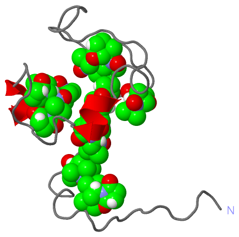
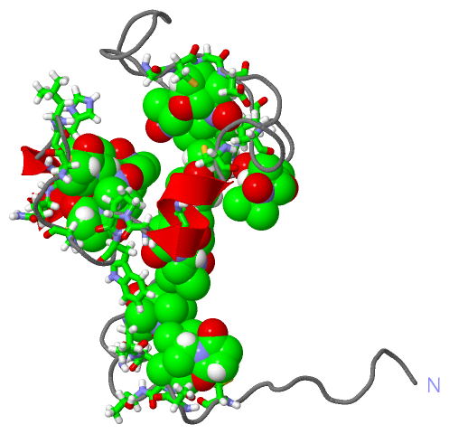
NMR Structure (1, 10)
No.NameCountTypeFull Name
1RCY10Ligand/Ion(3'R)-1'-OXYL-2',2',5',5'-TETRAMETHYL-1,3'-BIPYRROLIDINE-2,5-DIONE
NMR Structure * (1, 10)
No.NameCountTypeFull Name
1RCY10Ligand/Ion(3'R)-1'-OXYL-2',2',5',5'-TETRAMETHYL-1,3'-BIPYRROLIDINE-2,5-DIONE

(-) Sites  (10, 10)

NMR Structure (10, 10)
No.NameEvidenceResiduesDescription
01AC1SOFTWARECYS A:10 , SER A:12 , ALA A:13 , THR A:14BINDING SITE FOR RESIDUE RCY A 110
02AC2SOFTWAREVAL A:16 , MET A:17 , GLY A:18 , CYS A:21BINDING SITE FOR RESIDUE RCY A 121
03AC3SOFTWARECYS A:30 , ARG A:33BINDING SITE FOR RESIDUE RCY A 130
04AC4SOFTWAREARG A:33 , CYS A:38 , CYS A:50 , GLY A:51 , ASP A:52 , PRO A:55 , RCY A:150BINDING SITE FOR RESIDUE RCY A 138
05AC5SOFTWARECYS A:38 , GLY A:49 , CYS A:50 , GLY A:51 , ASP A:52 , RCY A:138BINDING SITE FOR RESIDUE RCY A 150
06AC6SOFTWAREPRO A:55 , CYS A:60 , ILE A:64 , RCY A:168BINDING SITE FOR RESIDUE RCY A 160
07AC7SOFTWAREILE A:64 , CYS A:68 , TRP A:70 , RCY A:160BINDING SITE FOR RESIDUE RCY A 168
08AC8SOFTWAREPRO A:69 , CYS A:73 , RCY A:176 , RCY A:187BINDING SITE FOR RESIDUE RCY A 173
09AC9SOFTWARECYS A:76 , LEU A:78 , HIS A:79 , ALA A:82 , GLN A:83 , RCY A:173 , RCY A:187BINDING SITE FOR RESIDUE RCY A 176
10BC1SOFTWARECYS A:87 , RCY A:173 , RCY A:176BINDING SITE FOR RESIDUE RCY A 187

(-) SS Bonds  (0, 0)

(no "SS Bond" information available for 2JU4)

(-) Cis Peptide Bonds  (0, 0)

(no "Cis Peptide Bond" information available for 2JU4)

 Sequence-Structure Mapping

(-) SAPs(SNPs)/Variants  (0, 0)

(no "SAP(SNP)/Variant" information available for 2JU4)

(-) PROSITE Motifs  (0, 0)

(no "PROSITE Motif" information available for 2JU4)

(-) Exons   (3, 3)

NMR Structure (3, 3)
 ENSEMBLUniProtKBPDB
No.Transcript IDExonExon IDGenome LocationLengthIDLocationLengthCountLocationLength
1.1ENSBTAT000000004591ENSBTAE00000003536chr19:52647788-52647987200CNRG_BOVIN1-61611A:1-6161
1.2ENSBTAT000000004592ENSBTAE00000003538chr19:52648443-5264848341CNRG_BOVIN61-75151A:61-7515
1.3ENSBTAT000000004593ENSBTAE00000003539chr19:52648922-52649512591CNRG_BOVIN75-99251A:75-8713

(-) Sequences/Alignments

NMR Structure
   Reformat: Number of residues per line =  ('0' or empty: single-line sequence representation)
  Number of residues per labelling interval =   
  UniProt sequence: complete  aligned part    
   Show mapping: SCOP domains CATH domains Pfam domains Secondary structure (by author)
SAPs(SNPs) PROSITE motifs Exons
(details for a mapped element are shown in a popup box when the mouse pointer rests over it)
Chain A from PDB  Type:PROTEIN  Length:87
 aligned with CNRG_BOVIN | P04972 from UniProtKB/Swiss-Prot  Length:87

    Alignment length:87
                                    10        20        30        40        50        60        70        80       
            CNRG_BOVIN    1 MNLEPPKAEIRSATRVMGGPVTPRKGPPKFKQRQTRQFKSKPPKKGVQGFGDDIPGMEGLGTDITVICPWEAFNHLELHELAQYGII 87
               SCOP domains --------------------------------------------------------------------------------------- SCOP domains
               CATH domains 2ju4A00 A:1-86 Intrinsically disordered gamma-subunit of cGMP phosphodiesterase       - CATH domains
               Pfam domains ----PDE6_gamma-2ju4A01 A:5-86                                                         - Pfam domains
         Sec.struct. author ............................................................hhhhhh.............hhhhh... Sec.struct. author
                 SAPs(SNPs) --------------------------------------------------------------------------------------- SAPs(SNPs)
                    PROSITE --------------------------------------------------------------------------------------- PROSITE
           Transcript 1 (1) Exon 1.1  PDB: A:1-61 UniProt: 1-61                          -------------Exon 1.3      Transcript 1 (1)
           Transcript 1 (2) ------------------------------------------------------------Exon 1.2       ------------ Transcript 1 (2)
                  2ju4 A  1 MNLEPPKAECRSATRVMGGPCTPRKGPPKCKQRQTRQCKSKPPKKGVQGCGDDIPGMEGCGTDITVICPWEACNHCELHELAQYGIC 87
                                    10        20        30        40        50        60        70        80       

   Legend:   → Mismatch (orange background)
  - → Gap (green background, '-', border residues have a numbering label)
    → Modified Residue (blue background, lower-case, 'x' indicates undefined single-letter code, labelled with number + name)
  x → Chemical Group (purple background, 'x', labelled with number + name, e.g. ACE or NH2)
  extra numbering lines below/above indicate numbering irregularities and modified residue names etc., number ends below/above '|'

 Classification and Annotation

(-) SCOP Domains  (0, 0)

(no "SCOP Domain" information available for 2JU4)

(-) CATH Domains  (1, 1)

NMR Structure

(-) Pfam Domains  (1, 1)

NMR Structure

(-) Gene Ontology  (11, 11)

NMR Structure(hide GO term definitions)
Chain A   (CNRG_BOVIN | P04972)
molecular function
    GO:0047555    3',5'-cyclic-GMP phosphodiesterase activity    Catalysis of the reaction: guanosine 3',5'-cyclic phosphate + H2O = guanosine 5'-phosphate.
    GO:0004114    3',5'-cyclic-nucleotide phosphodiesterase activity    Catalysis of the reaction: nucleoside 3',5'-cyclic phosphate + H2O = nucleoside 5'-phosphate.
    GO:0030553    cGMP binding    Interacting selectively and non-covalently with cGMP, the nucleotide cyclic GMP (guanosine 3',5'-cyclophosphate).
    GO:0016787    hydrolase activity    Catalysis of the hydrolysis of various bonds, e.g. C-O, C-N, C-C, phosphoric anhydride bonds, etc. Hydrolase is the systematic name for any enzyme of EC class 3.
biological process
    GO:0000187    activation of MAPK activity    The initiation of the activity of the inactive enzyme MAP kinase (MAPK).
    GO:0045745    positive regulation of G-protein coupled receptor protein signaling pathway    Any process that activates or increases the frequency, rate or extent of G-protein coupled receptor protein signaling pathway activity.
    GO:0045742    positive regulation of epidermal growth factor receptor signaling pathway    Any process that activates or increases the frequency, rate or extent of epidermal growth factor receptor signaling pathway activity.
    GO:0050896    response to stimulus    Any process that results in a change in state or activity of a cell or an organism (in terms of movement, secretion, enzyme production, gene expression, etc.) as a result of a stimulus. The process begins with detection of the stimulus and ends with a change in state or activity or the cell or organism.
    GO:0016056    rhodopsin mediated signaling pathway    The series of molecular signals generated as a consequence of excitation of rhodopsin by a photon and the events that convert the absorbed photons into a cellular response.
    GO:0007601    visual perception    The series of events required for an organism to receive a visual stimulus, convert it to a molecular signal, and recognize and characterize the signal. Visual stimuli are detected in the form of photons and are processed to form an image.
cellular component
    GO:0097381    photoreceptor disc membrane    Ovally-shaped membranous stack located inside the photoreceptor outer segment, and containing densely packed molecules of the photoreceptor protein rhodopsin that traverse the lipid bilayer. Disc membranes are apparently derived from the plasma membrane in the region of the cilium that connects the photoreceptor outer segment to the inner segment.

 Visualization

(-) Interactive Views

NMR Structure
  Complete Structure
    Jena3D(integrated viewing of ligand, site, SAP, PROSITE, SCOP information)
    WebMol | AstexViewer[tm]@PDBe
(Java Applets, require no local installation except for Java; loading may be slow)
    STRAP
(Java WebStart application, automatic local installation, requires Java; full application with system access!)
    RasMol
(require local installation)
    Molscript (VRML)
(requires installation of a VRML viewer; select preferred view via VRML and generate a mono or stereo PDF format file)
 
  Ligands, Modified Residues, Ions
    RCY  [ RasMol | Jena3D ]  +environment [ RasMol | Jena3D ]
 
  Sites
    AC1  [ RasMol ]  +environment [ RasMol ]
    AC2  [ RasMol ]  +environment [ RasMol ]
    AC3  [ RasMol ]  +environment [ RasMol ]
    AC4  [ RasMol ]  +environment [ RasMol ]
    AC5  [ RasMol ]  +environment [ RasMol ]
    AC6  [ RasMol ]  +environment [ RasMol ]
    AC7  [ RasMol ]  +environment [ RasMol ]
    AC8  [ RasMol ]  +environment [ RasMol ]
    AC9  [ RasMol ]  +environment [ RasMol ]
    BC1  [ RasMol ]  +environment [ RasMol ]
 
  Cis Peptide Bonds
(no "Cis Peptide Bonds" information available for 2ju4)
 

(-) Still Images

Jmol
  protein: cartoon or spacefill or dots and stick; nucleic acid: cartoon and stick; ligands: spacefill; active site: stick
Molscript
  protein, nucleic acid: cartoon; ligands: spacefill; active site: ball and stick

 Databases and Analysis Tools

(-) Databases

Access by PDB/NDB ID
  2ju4
    Family and Domain InformationProDom | SYSTERS
    General Structural InformationGlycoscienceDB | MMDB | NDB | OCA | PDB | PDBe | PDBj | PDBsum | PDBWiki | PQS | PROTEOPEDIA
    Orientation in MembranesOPM
    Protein SurfaceSURFACE
    Secondary StructureDSSP (structure derived) | HSSP (homology derived)
    Structural GenomicsGeneCensus
    Structural NeighboursCE | VAST
    Structure ClassificationCATH | Dali | SCOP
    Validation and Original DataBMRB Data View | BMRB Restraints Grid | EDS | PROCHECK | RECOORD | WHAT_CHECK
 
Access by UniProt ID/Accession number
  CNRG_BOVIN | P04972
    Comparative Protein Structure ModelsModBase
    Genomic InformationEnsembl
    Protein-protein InteractionDIP
    Sequence, Family and Domain InformationInterPro | Pfam | SMART | UniProtKB/SwissProt
 
Access by Enzyme Classificator   (EC Number)
  3.1.4.17
    General Enzyme InformationBRENDA | EC-PDB | Enzyme | IntEnz
    PathwayKEGG | MetaCyc
 
Access by Disease Identifier   (MIM ID)
  (no 'MIM ID' available)
    Disease InformationOMIM
 
Access by GenAge ID
  (no 'GenAge ID' available)
    Age Related InformationGenAge

(-) Analysis Tools

Access by PDB/NDB ID
    Domain InformationXDom
    Interatomic Contacts of Structural UnitsCSU
    Ligand-protein ContactsLPC
    Protein CavitiescastP
    Sequence and Secondary StructurePDBCartoon
    Structure AlignmentSTRAP(Java WebStart application, automatic local installation, requires Java; full application with system access!)
    Structure and Sequence BrowserSTING
 
Access by UniProt ID/Accession number
  CNRG_BOVIN | P04972
    Protein Disorder PredictionDisEMBL | FoldIndex | GLOBPLOT (for more information see DisProt)

 Related Entries

(-) Entries Sharing at Least One Protein Chain (UniProt ID)

UniProtKB/Swiss-Prot
        CNRG_BOVIN | P049721fqj 3jab 3jbq

(-) Related Entries Specified in the PDB File

(no "Related Entries Specified in the PDB File" available for 2JU4)