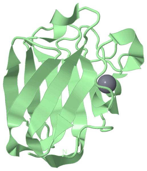
Show PDB file:   
         Plain Text   HTML   (compressed file size)
QuickSearch:   
by PDB,NDB,UniProt,PROSITE Code or Search Term(s)  
(-)Asymmetric Unit
(-)Asym. Unit - sites
(-)Biological Unit 1
(-)Biol. Unit 1 - sites
(-)Biological Unit 2
collapse expand < >
Image Asymmetric Unit
Asymmetric Unit  (Jmol Viewer)
Image Asym. Unit - sites
Asym. Unit - sites  (Jmol Viewer)
Image Biological Unit 1
Biological Unit 1  (Jmol Viewer)
Image Biol. Unit 1 - sites
Biol. Unit 1 - sites  (Jmol Viewer)
Image Biological Unit 2
Biological Unit 2  (Jmol Viewer)

(-) Description

Title :  STRUCTURE OF A STREPTOCOCCUS PNEUMONIAE FUCOSE BINDING MODULE
 
Authors :  A. B. Boraston, D. Wang, R. D. Burke
Date :  15 Aug 06  (Deposition) - 06 Sep 06  (Release) - 24 Feb 09  (Revision)
Method :  X-RAY DIFFRACTION
Resolution :  1.54
Chains :  Asym. Unit :  A,B
Biol. Unit 1:  A  (1x)
Biol. Unit 2:  B  (1x)
Keywords :  Sugar Binding Protein, Carbohydrate-Binding Protein (Keyword Search: [Gene Ontology, PubMed, Web (Google))
 
Reference :  A. B. Boraston, D. Wang, R. D. Burke
Blood Group Antigen Recognition By A Streptococcus Pneumoniae Virulence Factor.
J. Biol. Chem. V. 281 35263 2006
PubMed-ID: 16987809  |  Reference-DOI: 10.1074/JBC.M607620200

(-) Compounds

Molecule 1 - FUCOLECTIN-RELATED PROTEIN
    ChainsA, B
    EngineeredYES
    Expression SystemESCHERICHIA COLI
    Expression System Taxid562
    FragmentFUCOSE BINDING MODULE, RESIDUES 601-745
    Organism ScientificSTREPTOCOCCUS PNEUMONIAE
    Organism Taxid170187
    StrainTIGR4
    SynonymFUCOSE BINDING MODULE

 Structural Features

(-) Chains, Units

  12
Asymmetric Unit AB
Biological Unit 1 (1x)A 
Biological Unit 2 (1x) B

Summary Information (see also Sequences/Alignments below)

(-) Ligands, Modified Residues, Ions  (1, 2)

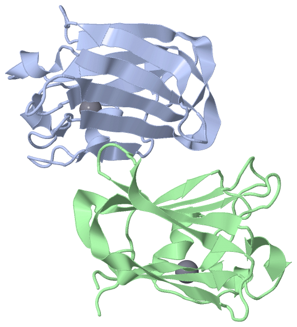
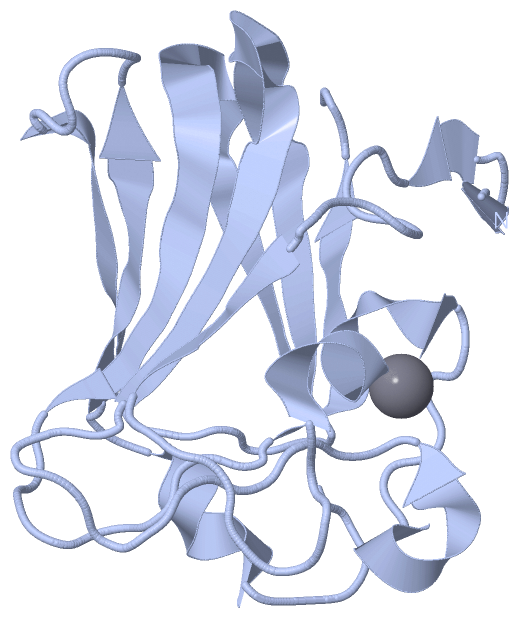
Asymmetric Unit (1, 2)
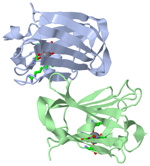
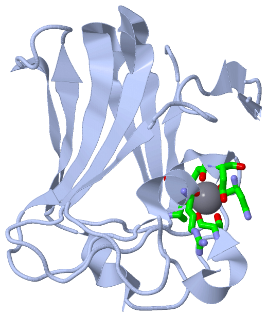
No.NameCountTypeFull Name
1CA2Ligand/IonCALCIUM ION
Biological Unit 1 (0, 0)
No.NameCountTypeFull Name
1CA-1Ligand/IonCALCIUM ION
Biological Unit 2 (0, 0)
No.NameCountTypeFull Name
1CA-1Ligand/IonCALCIUM ION

(-) Sites  (2, 2)

Asymmetric Unit (2, 2)
No.NameEvidenceResiduesDescription
1AC1SOFTWAREARG A:37 , ASP A:40 , ASN A:42 , SER A:51 , ALA A:143 , GLU A:144BINDING SITE FOR RESIDUE CA A1152
2AC2SOFTWAREARG B:37 , ASP B:40 , ASN B:42 , SER B:51 , ALA B:143 , GLU B:144BINDING SITE FOR RESIDUE CA B1152

(-) SS Bonds  (0, 0)

(no "SS Bond" information available for 2J1R)

(-) Cis Peptide Bonds  (2, 2)

Asymmetric Unit
No.Residues
1Asn A:59 -Pro A:60
2Asn B:59 -Pro B:60

 Sequence-Structure Mapping

(-) SAPs(SNPs)/Variants  (0, 0)

(no "SAP(SNP)/Variant" information available for 2J1R)

(-) PROSITE Motifs  (0, 0)

(no "PROSITE Motif" information available for 2J1R)

(-) Exons   (0, 0)

(no "Exon" information available for 2J1R)

(-) Sequences/Alignments

Asymmetric Unit
   Reformat: Number of residues per line =  ('0' or empty: single-line sequence representation)
  Number of residues per labelling interval =   
  UniProt sequence: complete  aligned part    
   Show mapping: SCOP domains CATH domains Pfam domains Secondary structure (by author)
SAPs(SNPs) PROSITE motifs Exons
(details for a mapped element are shown in a popup box when the mouse pointer rests over it)
Chain A from PDB  Type:PROTEIN  Length:142
 aligned with A0A0H2US34_S | A0A0H2US34 from UniProtKB/TrEMBL  Length:1038

    Alignment length:142
                                   613       623       633       643       653       663       673       683       693       703       713       723       733       743  
         A0A0H2US34_S   604 KFNDGNLNIAYAKPTTQSSVDYNGDPNRAVDGNRNGNFNSGSVTHTRADNPSWWEVDLKKMDKVGLVKIYNRTDAETQRLSNFDVILYDNNRNEVAKKHVNNLSGESVSLDFKEKGARYIKVKLLTSGVPLSLAEVEVFRES 745
               SCOP domains d2j1ra_ A: automated matches                                                                                                                   SCOP domains
               CATH domains ---------------------------------------------------------------------------------------------------------------------------------------------- CATH domains
               Pfam domains ---------------------------------------------------------------------------------------------------------------------------------------------- Pfam domains
         Sec.struct. author .ee..ee.......eee.......hhhhhhh.....hhhhh...........eeeeeeeeeeeeeeeeeee....hhhhh..eeeeee.....eeeeeee......eeeeeeeeeeeeeeeeee........eeeeee.... Sec.struct. author
                 SAPs(SNPs) ---------------------------------------------------------------------------------------------------------------------------------------------- SAPs(SNPs)
                    PROSITE ---------------------------------------------------------------------------------------------------------------------------------------------- PROSITE
                 Transcript ---------------------------------------------------------------------------------------------------------------------------------------------- Transcript
                 2j1r A  10 KFNDGNLNIAYAKPTTQSSVDYNGDPNRAVDGNRNGNFNSGSVTHTRADNPSWWEVDLKKMDKVGLVKIYNRTDAETQRLSNFDVILYDNNRNEVAKKHVNNLSGESVSLDFKEKGARYIKVKLLTSGVPLSLAEVEVFRES 151
                                    19        29        39        49        59        69        79        89        99       109       119       129       139       149  

Chain B from PDB  Type:PROTEIN  Length:137
 aligned with A0A0H2US34_S | A0A0H2US34 from UniProtKB/TrEMBL  Length:1038

    Alignment length:137
                                   618       628       638       648       658       668       678       688       698       708       718       728       738       
         A0A0H2US34_S   609 NLNIAYAKPTTQSSVDYNGDPNRAVDGNRNGNFNSGSVTHTRADNPSWWEVDLKKMDKVGLVKIYNRTDAETQRLSNFDVILYDNNRNEVAKKHVNNLSGESVSLDFKEKGARYIKVKLLTSGVPLSLAEVEVFRES 745
               SCOP domains d2j1rb_ B: automated matches                                                                                                              SCOP domains
               CATH domains ----------------------------------------------------------------------------------------------------------------------------------------- CATH domains
               Pfam domains ----------------------------------------------------------------------------------------------------------------------------------------- Pfam domains
         Sec.struct. author .........eee.......hhhhhhh.....hhhhh...........eeeeeeeeeeeeeeeeeee....hhhhh..eeeeee.....eeeeeee......eeeeeeeeeeeeeeeeee........eeeeee.... Sec.struct. author
                 SAPs(SNPs) ----------------------------------------------------------------------------------------------------------------------------------------- SAPs(SNPs)
                    PROSITE ----------------------------------------------------------------------------------------------------------------------------------------- PROSITE
                 Transcript ----------------------------------------------------------------------------------------------------------------------------------------- Transcript
                 2j1r B  15 NLNIAYAKPTTQSSVDYNGDPNRAVDGNRNGNFNSGSVTHTRADNPSWWEVDLKKMDKVGLVKIYNRTDAETQRLSNFDVILYDNNRNEVAKKHVNNLSGESVSLDFKEKGARYIKVKLLTSGVPLSLAEVEVFRES 151
                                    24        34        44        54        64        74        84        94       104       114       124       134       144       

   Legend:   → Mismatch (orange background)
  - → Gap (green background, '-', border residues have a numbering label)
    → Modified Residue (blue background, lower-case, 'x' indicates undefined single-letter code, labelled with number + name)
  x → Chemical Group (purple background, 'x', labelled with number + name, e.g. ACE or NH2)
  extra numbering lines below/above indicate numbering irregularities and modified residue names etc., number ends below/above '|'

 Classification and Annotation

(-) SCOP Domains  (1, 2)

Asymmetric Unit

(-) CATH Domains  (0, 0)

(no "CATH Domain" information available for 2J1R)

(-) Pfam Domains  (0, 0)

(no "Pfam Domain" information available for 2J1R)

(-) Gene Ontology  (0, 0)

Asymmetric Unit(hide GO term definitions)
    (no "Gene Ontology" information available for 2J1R)

 Visualization

(-) Interactive Views

Asymmetric Unit
  Complete Structure
    Jena3D(integrated viewing of ligand, site, SAP, PROSITE, SCOP information)
    WebMol | AstexViewer[tm]@PDBe
(Java Applets, require no local installation except for Java; loading may be slow)
    STRAP
(Java WebStart application, automatic local installation, requires Java; full application with system access!)
    RasMol
(require local installation)
    Molscript (VRML)
(requires installation of a VRML viewer; select preferred view via VRML and generate a mono or stereo PDF format file)
 
  Ligands, Modified Residues, Ions
    CA  [ RasMol | Jena3D ]  +environment [ RasMol | Jena3D ]
 
  Sites
    AC1  [ RasMol ]  +environment [ RasMol ]
    AC2  [ RasMol ]  +environment [ RasMol ]
 
  Cis Peptide Bonds
    Asn A:59 - Pro A:60   [ RasMol ]  
    Asn B:59 - Pro B:60   [ RasMol ]  
 
Biological Units
  Complete Structure
    Biological Unit 1  [ Jena3D ]
    Biological Unit 2  [ Jena3D ]

(-) Still Images

Jmol
  protein: cartoon or spacefill or dots and stick; nucleic acid: cartoon and stick; ligands: spacefill; active site: stick
Molscript
  protein, nucleic acid: cartoon; ligands: spacefill; active site: ball and stick

 Databases and Analysis Tools

(-) Databases

Access by PDB/NDB ID
  2j1r
    Family and Domain InformationProDom | SYSTERS
    General Structural InformationGlycoscienceDB | MMDB | NDB | OCA | PDB | PDBe | PDBj | PDBsum | PDBWiki | PQS | PROTEOPEDIA
    Orientation in MembranesOPM
    Protein SurfaceSURFACE
    Secondary StructureDSSP (structure derived) | HSSP (homology derived)
    Structural GenomicsGeneCensus
    Structural NeighboursCE | VAST
    Structure ClassificationCATH | Dali | SCOP
    Validation and Original DataBMRB Data View | BMRB Restraints Grid | EDS | PROCHECK | RECOORD | WHAT_CHECK
 
Access by UniProt ID/Accession number
  A0A0H2US34_S | A0A0H2US34
    Comparative Protein Structure ModelsModBase
    Genomic InformationEnsembl
    Protein-protein InteractionDIP
    Sequence, Family and Domain InformationInterPro | Pfam | SMART | UniProtKB/TrEMBL
 
Access by Enzyme Classificator   (EC Number)
  (no 'Enzyme Classificator' available)
    General Enzyme InformationBRENDA | EC-PDB | Enzyme | IntEnz
    PathwayKEGG | MetaCyc
 
Access by Disease Identifier   (MIM ID)
  (no 'MIM ID' available)
    Disease InformationOMIM
 
Access by GenAge ID
  (no 'GenAge ID' available)
    Age Related InformationGenAge

(-) Analysis Tools

Access by PDB/NDB ID
    Domain InformationXDom
    Interatomic Contacts of Structural UnitsCSU
    Ligand-protein ContactsLPC
    Protein CavitiescastP
    Sequence and Secondary StructurePDBCartoon
    Structure AlignmentSTRAP(Java WebStart application, automatic local installation, requires Java; full application with system access!)
    Structure and Sequence BrowserSTING
 
Access by UniProt ID/Accession number
  A0A0H2US34_S | A0A0H2US34
    Protein Disorder PredictionDisEMBL | FoldIndex | GLOBPLOT (for more information see DisProt)

 Related Entries

(-) Entries Sharing at Least One Protein Chain (UniProt ID)

UniProtKB/TrEMBL
        A0A0H2US34_S | A0A0H2US342j1s 2j1t 2j1u 2j1v 2j22 2wmf 2wmg 2wmh

(-) Related Entries Specified in the PDB File

2j1s STRUCTURE OF A STREPTOCOCCUS PNEUMONIAE FUCOSE BINDING MODULE IN COMPLEX WITH FUCOSE
2j1t STRUCTURE OF A STREPTOCOCCUS PNEUMONIAE FUCOSE BINDING MODULE IN COMPLEX WITH THE LEWIS Y ANTIGEN
2j1u STRUCTURE OF A STREPTOCOCCUS PNEUMONIAE FUCOSE BINDING MODULE IN COMPLEX WITH THE BLOOD GROUP A-TETRASACCHARIDE
2j1v STRUCTURE OF A STREPTOCOCCUS PNEUMONIAE FUCOSE BINDING MODULE IN COMPLEX WITH THE BLOOD GROUP H-TRISACCHARIDE
2j22 STRUCTURE OF A STREPTOCOCCUS PNEUMONIAE FUCOSE BINDING MODULE, SPX-3