| molecular function |
|---|
| | GO:0005518 | | collagen binding | | Interacting selectively and non-covalently with collagen, a group of fibrous proteins of very high tensile strength that form the main component of connective tissue in animals. Collagen is highly enriched in glycine (some regions are 33% glycine) and proline, occurring predominantly as 3-hydroxyproline (about 20%). |
| | GO:0004197 | | cysteine-type endopeptidase activity | | Catalysis of the hydrolysis of internal, alpha-peptide bonds in a polypeptide chain by a mechanism in which the sulfhydryl group of a cysteine residue at the active center acts as a nucleophile. |
| | GO:0008234 | | cysteine-type peptidase activity | | Catalysis of the hydrolysis of peptide bonds in a polypeptide chain by a mechanism in which the sulfhydryl group of a cysteine residue at the active center acts as a nucleophile. |
| | GO:0001968 | | fibronectin binding | | Interacting selectively and non-covalently with a fibronectin, a group of related adhesive glycoproteins of high molecular weight found on the surface of animal cells, connective tissue matrices, and in extracellular fluids. |
| | GO:0016787 | | hydrolase activity | | Catalysis of the hydrolysis of various bonds, e.g. C-O, C-N, C-C, phosphoric anhydride bonds, etc. Hydrolase is the systematic name for any enzyme of EC class 3. |
| | GO:0043236 | | laminin binding | | Interacting selectively and non-covalently with laminins, glycoproteins that are major constituents of the basement membrane of cells. |
| | GO:0008233 | | peptidase activity | | Catalysis of the hydrolysis of a peptide bond. A peptide bond is a covalent bond formed when the carbon atom from the carboxyl group of one amino acid shares electrons with the nitrogen atom from the amino group of a second amino acid. |
| | GO:0043394 | | proteoglycan binding | | Interacting selectively and non-covalently with a proteoglycan, any glycoprotein in which the carbohydrate units are glycosaminoglycans. |
| | GO:0004252 | | serine-type endopeptidase activity | | Catalysis of the hydrolysis of internal, alpha-peptide bonds in a polypeptide chain by a catalytic mechanism that involves a catalytic triad consisting of a serine nucleophile that is activated by a proton relay involving an acidic residue (e.g. aspartate or glutamate) and a basic residue (usually histidine). |
| biological process |
|---|
| | GO:0002250 | | adaptive immune response | | An immune response mediated by cells expressing specific receptors for antigen produced through a somatic diversification process, and allowing for an enhanced secondary response to subsequent exposures to the same antigen (immunological memory). |
| | GO:0019882 | | antigen processing and presentation | | The process in which an antigen-presenting cell expresses antigen (peptide or lipid) on its cell surface in association with an MHC protein complex. |
| | GO:0019886 | | antigen processing and presentation of exogenous peptide antigen via MHC class II | | The process in which an antigen-presenting cell expresses a peptide antigen of exogenous origin on its cell surface in association with an MHC class II protein complex. The peptide antigen is typically, but not always, processed from a whole protein. |
| | GO:0048002 | | antigen processing and presentation of peptide antigen | | The process in which an antigen-presenting cell expresses peptide antigen in association with an MHC protein complex on its cell surface, including proteolysis and transport steps for the peptide antigen both prior to and following assembly with the MHC protein complex. The peptide antigen is typically, but not always, processed from an endogenous or exogenous protein. |
| | GO:0034769 | | basement membrane disassembly | | The controlled breakdown of the basement membrane in the context of a normal process such as imaginal disc eversion. |
| | GO:0097067 | | cellular response to thyroid hormone stimulus | | A change in state or activity of a cell (in terms of movement, secretion, enzyme production, gene expression, etc.) as a result of a thyroid hormone stimulus. |
| | GO:0030574 | | collagen catabolic process | | The proteolytic chemical reactions and pathways resulting in the breakdown of collagen in the extracellular matrix, usually carried out by proteases secreted by nearby cells. |
| | GO:0022617 | | extracellular matrix disassembly | | A process that results in the breakdown of the extracellular matrix. |
| | GO:0006955 | | immune response | | Any immune system process that functions in the calibrated response of an organism to a potential internal or invasive threat. |
| | GO:2001259 | | positive regulation of cation channel activity | | Any process that activates or increases the frequency, rate or extent of cation channel activity. |
| | GO:0016485 | | protein processing | | Any protein maturation process achieved by the cleavage of a peptide bond or bonds within a protein. Protein maturation is the process leading to the attainment of the full functional capacity of a protein. |
| | GO:0006508 | | proteolysis | | The hydrolysis of proteins into smaller polypeptides and/or amino acids by cleavage of their peptide bonds. |
| | GO:0051603 | | proteolysis involved in cellular protein catabolic process | | The hydrolysis of a peptide bond or bonds within a protein as part of the chemical reactions and pathways resulting in the breakdown of a protein by individual cells. |
| | GO:0010447 | | response to acidic pH | | Any process that results in a change in state or activity of a cell or an organism (in terms of movement, secretion, enzyme production, gene expression, etc.) as a result of a pH stimulus with pH < 7. pH is a measure of the acidity or basicity of an aqueous solution. |
| | GO:0002224 | | toll-like receptor signaling pathway | | Any series of molecular signals generated as a consequence of binding to a toll-like receptor. Toll-like receptors directly bind pattern motifs from a variety of microbial sources to initiate innate immune response. |
| cellular component |
|---|
| | GO:0036021 | | endolysosome lumen | | The volume enclosed by the membrane of an endolysosome. An endolysosome is a transient hybrid organelle formed by fusion of a late endosome with a lysosome. |
| | GO:0005576 | | extracellular region | | The space external to the outermost structure of a cell. For cells without external protective or external encapsulating structures this refers to space outside of the plasma membrane. This term covers the host cell environment outside an intracellular parasite. |
| | GO:0005615 | | extracellular space | | That part of a multicellular organism outside the cells proper, usually taken to be outside the plasma membranes, and occupied by fluid. |
| | GO:0043231 | | intracellular membrane-bounded organelle | | Organized structure of distinctive morphology and function, bounded by a single or double lipid bilayer membrane and occurring within the cell. Includes the nucleus, mitochondria, plastids, vacuoles, and vesicles. Excludes the plasma membrane. |
| | GO:0043202 | | lysosomal lumen | | The volume enclosed within the lysosomal membrane. |
| | GO:0005764 | | lysosome | | A small lytic vacuole that has cell cycle-independent morphology and is found in most animal cells and that contains a variety of hydrolases, most of which have their maximal activities in the pH range 5-6. The contained enzymes display latency if properly isolated. About 40 different lysosomal hydrolases are known and lysosomes have a great variety of morphologies and functions. |
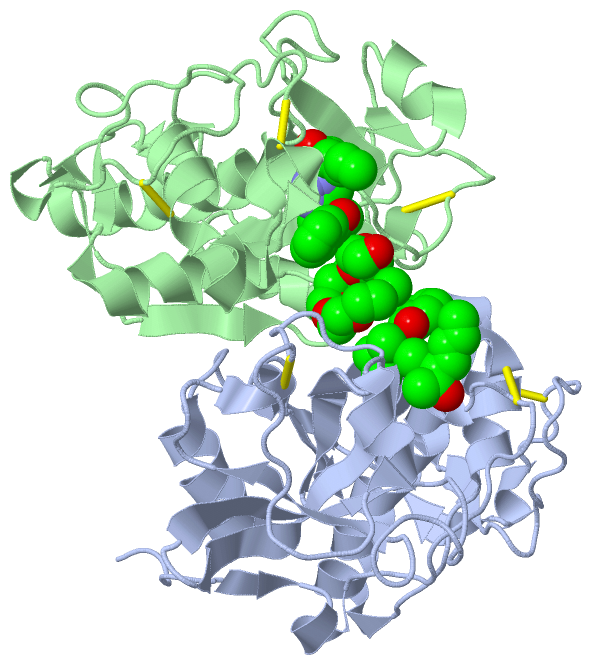
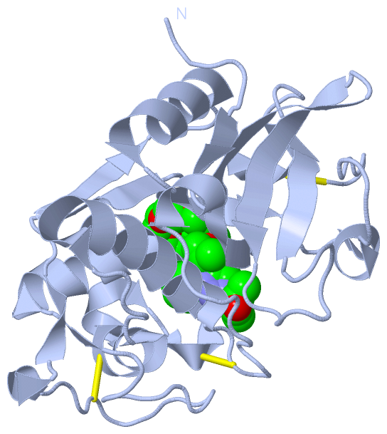
 Description
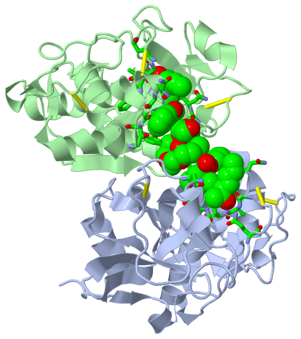
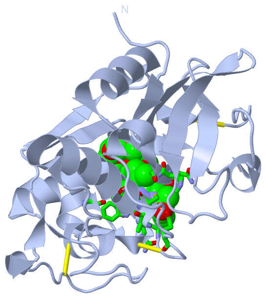