Show PDB file:   
         Plain Text   HTML   (compressed file size)
QuickSearch:   
by PDB,NDB,UniProt,PROSITE Code or Search Term(s)  
(-)Asymmetric Unit
(-)Asym. Unit - sites
(-)Biological Unit 1
(-)Biol. Unit 1 - sites
(-)Biological Unit 2
(-)Biological Unit 3
collapse expand < >
Image Asymmetric Unit
Asymmetric Unit  (Jmol Viewer)
Image Asym. Unit - sites
Asym. Unit - sites  (Jmol Viewer)
Image Biological Unit 1
Biological Unit 1  (Jmol Viewer)
Image Biol. Unit 1 - sites
Biol. Unit 1 - sites  (Jmol Viewer)
Image Biological Unit 2
Biological Unit 2  (Jmol Viewer)
Image Biological Unit 3
Biological Unit 3  (Jmol Viewer)

(-) Description

Title :  CRYSTAL STRUCTURE OF HUMAN CATHEPSIN S BOUND TO A NON-COVALENT INHIBITOR
 
Authors :  Y. Wang, P. K. Jadhav, G. G. Deng
Date :  24 Mar 14  (Deposition) - 29 Oct 14  (Release) - 29 Oct 14  (Revision)
Method :  X-RAY DIFFRACTION
Resolution :  1.80
Chains :  Asym. Unit :  A,B
Biol. Unit 1:  A  (1x)
Biol. Unit 2:  B  (1x)
Biol. Unit 3:  A (1x),B (1x)
Keywords :  Cysteine Protease, Cathepsin S, Inhibitor, Non-Covalent (Keyword Search: [Gene Ontology, PubMed, Web (Google))
 
Reference :  P. K. Jadhav, M. A. Schiffler, K. Gavardinas, E. J. Kim, D. P. Matthews, M. A. Staszak, D. S. Coffey, B. W. Shaw, K. C. Cassidy, R. A. Brier, Y. Zhang, R. M. Christie, W. F. Matter, K. Qing, J. D. Durbin, Y. Wang, G. G. Deng
Discovery Of Cathepsin S Inhibitor Ly3000328 For The Treatment Of Abdominal Aortic Aneurysm.
Acs Med. Chem. Lett. V. 5 1138 2014
PubMed-ID: 25313327  |  Reference-DOI: 10.1021/ML500283G

(-) Compounds

Molecule 1 - CATHEPSIN S
    ChainsA, B
    EC Number3.4.22.27
    EngineeredYES
    Expression SystemENTEROBACTERIA PHAGE L1
    Expression System Taxid268588
    GeneCTSS
    Organism CommonHUMAN
    Organism ScientificHOMO SAPIENS
    Organism Taxid9606

 Structural Features

(-) Chains, Units

  12
Asymmetric Unit AB
Biological Unit 1 (1x)A 
Biological Unit 2 (1x) B
Biological Unit 3 (1x)A (1x)B (1x)

Summary Information (see also Sequences/Alignments below)

(-) Ligands, Modified Residues, Ions  (2, 3)

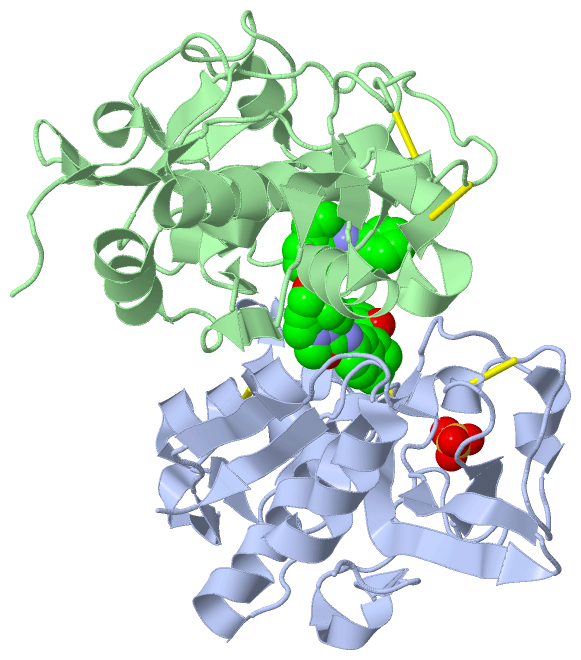
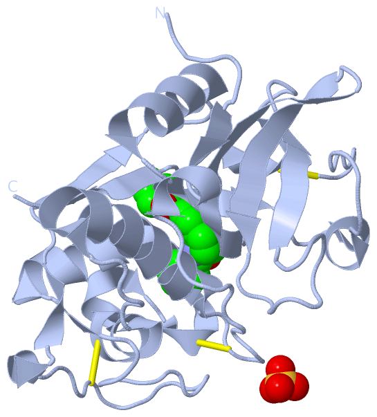
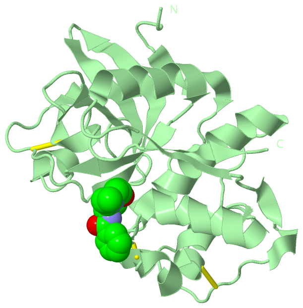
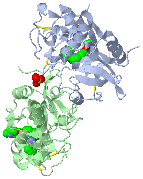
Asymmetric Unit (2, 3)
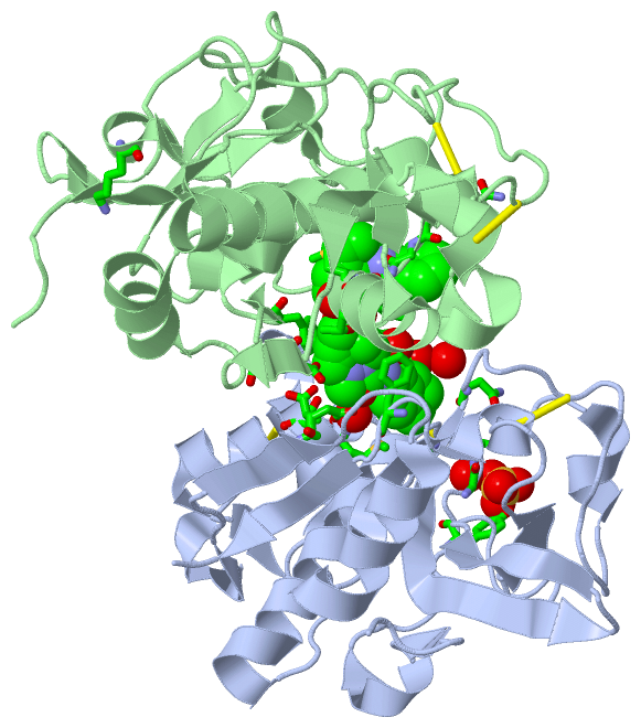
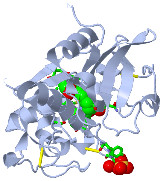
No.NameCountTypeFull Name
12FC2Ligand/IonN-[(8R)-8-(BENZOYLAMINO)-5,6,7,8-TETRAHYDRONAPHTHALEN-2-YL]-4-METHYLPIPERAZINE-1-CARBOXAMIDE
2SO41Ligand/IonSULFATE ION
Biological Unit 1 (2, 2)
No.NameCountTypeFull Name
12FC1Ligand/IonN-[(8R)-8-(BENZOYLAMINO)-5,6,7,8-TETRAHYDRONAPHTHALEN-2-YL]-4-METHYLPIPERAZINE-1-CARBOXAMIDE
2SO41Ligand/IonSULFATE ION
Biological Unit 2 (1, 1)
No.NameCountTypeFull Name
12FC1Ligand/IonN-[(8R)-8-(BENZOYLAMINO)-5,6,7,8-TETRAHYDRONAPHTHALEN-2-YL]-4-METHYLPIPERAZINE-1-CARBOXAMIDE
2SO4-1Ligand/IonSULFATE ION
Biological Unit 3 (2, 2)
No.NameCountTypeFull Name
12FC1Ligand/IonN-[(8R)-8-(BENZOYLAMINO)-5,6,7,8-TETRAHYDRONAPHTHALEN-2-YL]-4-METHYLPIPERAZINE-1-CARBOXAMIDE
2SO41Ligand/IonSULFATE ION

(-) Sites  (3, 3)

Asymmetric Unit (3, 3)
No.NameEvidenceResiduesDescription
1AC1SOFTWARETYR A:18 , GLY A:20 , SER A:21 , HOH A:530 , LYS B:10binding site for residue SO4 A 301
2AC2SOFTWAREGLY A:62 , GLY A:68 , GLY A:69 , PHE A:70 , MET A:71 , ASN A:163 , GLY A:165 , PHE A:211 , HOH A:469 , HOH A:578 , HOH A:593 , PHE B:70 , THR B:72 , 2FC B:301binding site for residue 2FC A 302
3AC3SOFTWAREPHE A:70 , THR A:72 , GLU A:115 , 2FC A:302 , GLY B:62 , ASN B:67 , GLY B:68 , GLY B:69 , PHE B:70 , MET B:71 , GLU B:115 , ASN B:163 , GLY B:165 , PHE B:211 , HOH B:451 , HOH B:592binding site for residue 2FC B 301

(-) SS Bonds  (6, 6)

Asymmetric Unit
No.Residues
1A:22 -A:66
2A:56 -A:99
3A:158 -A:206
4B:22 -B:66
5B:56 -B:99
6B:158 -B:206

(-) Cis Peptide Bonds  (0, 0)

(no "Cis Peptide Bond" information available for 4P6E)

 Sequence-Structure Mapping

(-) SAPs(SNPs)/Variants  (0, 0)

(no "SAP(SNP)/Variant" information available for 4P6E)

(-) PROSITE Motifs  (0, 0)

(no "PROSITE Motif" information available for 4P6E)

(-) Exons   (0, 0)

(no "Exon" information available for 4P6E)

(-) Sequences/Alignments

Asymmetric Unit
   Reformat: Number of residues per line =  ('0' or empty: single-line sequence representation)
  Number of residues per labelling interval =   
  UniProt sequence: complete  aligned part    
   Show mapping: SCOP domains CATH domains Pfam domains Secondary structure (by author)
SAPs(SNPs) PROSITE motifs Exons
(details for a mapped element are shown in a popup box when the mouse pointer rests over it)
Chain A from PDB  Type:PROTEIN  Length:219
                                                                                                                                                                                                                                                           
               SCOP domains --------------------------------------------------------------------------------------------------------------------------------------------------------------------------------------------------------------------------- SCOP domains
               CATH domains --------------------------------------------------------------------------------------------------------------------------------------------------------------------------------------------------------------------------- CATH domains
               Pfam domains --------------------------------------------------------------------------------------------------------------------------------------------------------------------------------------------------------------------------- Pfam domains
         Sec.struct. author .....ee.hhhh............hhhhhhhhhhhhhhhhhhhh.....hhhhhhhhhhhhhh.hhhhh.hhhhhhhhhhhhh.ee................hhh.eee...eeee....hhhhhhhhhhhh..eeeee...hhhhhh................eeeeeeeeeee..eeeeeee...........eeeee....hhhhh....eeee.. Sec.struct. author
                 SAPs(SNPs) --------------------------------------------------------------------------------------------------------------------------------------------------------------------------------------------------------------------------- SAPs(SNPs)
                    PROSITE --------------------------------------------------------------------------------------------------------------------------------------------------------------------------------------------------------------------------- PROSITE
                 Transcript --------------------------------------------------------------------------------------------------------------------------------------------------------------------------------------------------------------------------- Transcript
                 4p6e A   0 ILPDSVDWREKGCVTEVKYQGSCGACWAFSAVGALEAQLKLKTGKLVSLSAQNLVDCSTEKYGNKGCNGGFMTTAFQYIIDNKGIDSDASYPYKAMDQKCQYDSKYRAATCSKYTELPYGREDVLKEAVANKGPVSVGVDARHPSFFLYRSGVYYEPSCTQNVNHGVLVVGYGDLNGKEYWLVKNSWGHNFGEEGYIRMARNKGNHCGIASFPSYPEIE 218
                                     9        19        29        39        49        59        69        79        89        99       109       119       129       139       149       159       169       179       189       199       209         

Chain B from PDB  Type:PROTEIN  Length:219
                                                                                                                                                                                                                                                           
               SCOP domains --------------------------------------------------------------------------------------------------------------------------------------------------------------------------------------------------------------------------- SCOP domains
               CATH domains --------------------------------------------------------------------------------------------------------------------------------------------------------------------------------------------------------------------------- CATH domains
               Pfam domains --------------------------------------------------------------------------------------------------------------------------------------------------------------------------------------------------------------------------- Pfam domains
         Sec.struct. author .....ee.hhhh............hhhhhhhhhhhhhhhhhhhh.....hhhhhhhhhhhhhh.hhhhh.hhhhhhhhhhhhh.ee................hhh.eee...eeee....hhhhhhhhhhhh..eeeee...hhhhhh....ee..........eeeeeeeeeee..eeeeeee...........eeeee....hhhhh....eeee.. Sec.struct. author
                 SAPs(SNPs) --------------------------------------------------------------------------------------------------------------------------------------------------------------------------------------------------------------------------- SAPs(SNPs)
                    PROSITE --------------------------------------------------------------------------------------------------------------------------------------------------------------------------------------------------------------------------- PROSITE
                 Transcript --------------------------------------------------------------------------------------------------------------------------------------------------------------------------------------------------------------------------- Transcript
                 4p6e B   0 ILPDSVDWREKGCVTEVKYQGSCGACWAFSAVGALEAQLKLKTGKLVSLSAQNLVDCSTEKYGNKGCNGGFMTTAFQYIIDNKGIDSDASYPYKAMDQKCQYDSKYRAATCSKYTELPYGREDVLKEAVANKGPVSVGVDARHPSFFLYRSGVYYEPSCTQNVNHGVLVVGYGDLNGKEYWLVKNSWGHNFGEEGYIRMARNKGNHCGIASFPSYPEIE 218
                                     9        19        29        39        49        59        69        79        89        99       109       119       129       139       149       159       169       179       189       199       209         

   Legend:   → Mismatch (orange background)
  - → Gap (green background, '-', border residues have a numbering label)
    → Modified Residue (blue background, lower-case, 'x' indicates undefined single-letter code, labelled with number + name)
  x → Chemical Group (purple background, 'x', labelled with number + name, e.g. ACE or NH2)
  extra numbering lines below/above indicate numbering irregularities and modified residue names etc., number ends below/above '|'

 Classification and Annotation

(-) SCOP Domains  (0, 0)

(no "SCOP Domain" information available for 4P6E)

(-) CATH Domains  (0, 0)

(no "CATH Domain" information available for 4P6E)

(-) Pfam Domains  (0, 0)

(no "Pfam Domain" information available for 4P6E)

(-) Gene Ontology  (30, 30)

Asymmetric Unit(hide GO term definitions)

 Visualization

(-) Interactive Views

Asymmetric Unit
  Complete Structure
    Jena3D(integrated viewing of ligand, site, SAP, PROSITE, SCOP information)
    WebMol | AstexViewer[tm]@PDBe
(Java Applets, require no local installation except for Java; loading may be slow)
    STRAP
(Java WebStart application, automatic local installation, requires Java; full application with system access!)
    RasMol
(require local installation)
    Molscript (VRML)
(requires installation of a VRML viewer; select preferred view via VRML and generate a mono or stereo PDF format file)
 
  Ligands, Modified Residues, Ions
    2FC  [ RasMol | Jena3D ]  +environment [ RasMol | Jena3D ]
    SO4  [ RasMol | Jena3D ]  +environment [ RasMol | Jena3D ]
 
  Sites
    AC1  [ RasMol ]  +environment [ RasMol ]
    AC2  [ RasMol ]  +environment [ RasMol ]
    AC3  [ RasMol ]  +environment [ RasMol ]
 
  Cis Peptide Bonds
(no "Cis Peptide Bonds" information available for 4p6e)
 
Biological Units
  Complete Structure
    Biological Unit 1  [ Jena3D ]
    Biological Unit 2  [ Jena3D ]
    Biological Unit 3  [ Jena3D ]

(-) Still Images

Jmol
  protein: cartoon or spacefill or dots and stick; nucleic acid: cartoon and stick; ligands: spacefill; active site: stick
Molscript
  protein, nucleic acid: cartoon; ligands: spacefill; active site: ball and stick

 Databases and Analysis Tools

(-) Databases

Access by PDB/NDB ID
  4p6e
    Family and Domain InformationProDom | SYSTERS
    General Structural InformationGlycoscienceDB | MMDB | NDB | OCA | PDB | PDBe | PDBj | PDBsum | PDBWiki | PQS | PROTEOPEDIA
    Orientation in MembranesOPM
    Protein SurfaceSURFACE
    Secondary StructureDSSP (structure derived) | HSSP (homology derived)
    Structural GenomicsGeneCensus
    Structural NeighboursCE | VAST
    Structure ClassificationCATH | Dali | SCOP
    Validation and Original DataBMRB Data View | BMRB Restraints Grid | EDS | PROCHECK | RECOORD | WHAT_CHECK
 
Access by UniProt ID/Accession number
  CATS_HUMAN | P25774
    Comparative Protein Structure ModelsModBase
    Genomic InformationEnsembl
    Protein-protein InteractionDIP
    Sequence, Family and Domain InformationInterPro | Pfam | SMART | UniProtKB/SwissProt
 
Access by Enzyme Classificator   (EC Number)
  3.4.22.27
    General Enzyme InformationBRENDA | EC-PDB | Enzyme | IntEnz
    PathwayKEGG | MetaCyc
 
Access by Disease Identifier   (MIM ID)
  (no 'MIM ID' available)
    Disease InformationOMIM
 
Access by GenAge ID
  (no 'GenAge ID' available)
    Age Related InformationGenAge

(-) Analysis Tools

Access by PDB/NDB ID
    Domain InformationXDom
    Interatomic Contacts of Structural UnitsCSU
    Ligand-protein ContactsLPC
    Protein CavitiescastP
    Sequence and Secondary StructurePDBCartoon
    Structure AlignmentSTRAP(Java WebStart application, automatic local installation, requires Java; full application with system access!)
    Structure and Sequence BrowserSTING
 
Access by UniProt ID/Accession number
  CATS_HUMAN | P25774
    Protein Disorder PredictionDisEMBL | FoldIndex | GLOBPLOT (for more information see DisProt)

 Related Entries

(-) Entries Sharing at Least One Protein Chain (UniProt ID)

UniProtKB/Swiss-Prot
        CATS_HUMAN | P257741bxf 1glo 1ms6 1npz 1nqc 2c0y 2f1g 2fq9 2fra 2frq 2ft2 2fud 2fye 2g6d 2g7y 2h7j 2hh5 2hhn 2hxz 2op3 2r9m 2r9n 2r9o 3iej 3kwn 3mpe 3mpf 3n3g 3n4c 3ovx 4p6g

(-) Related Entries Specified in the PDB File

(no "Related Entries Specified in the PDB File" available for 4P6E)