Show PDB file:   
         Plain Text   HTML   (compressed file size)
QuickSearch:   
by PDB,NDB,UniProt,PROSITE Code or Search Term(s)  
(-)Asym./Biol. Unit - manually
(-)Asym./Biol. Unit
collapse expand < >
Image Asym./Biol. Unit - manually
Asym./Biol. Unit - manually  (Jmol Viewer)
Image Asym./Biol. Unit
Asym./Biol. Unit  (Jmol Viewer)

(-) Description

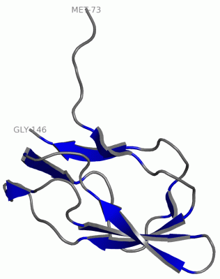
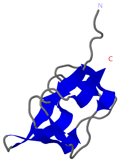
Title :  STRUCTURE OF BIOTIN CARBOXYL CARRIER PROTEIN (74VAL START) FROM PYROCOCCUS HORIKOSHI OT3 LIGAND FREE FORM I
 
Authors :  B. Bagautdinov, N. Kunishima, Riken Structural Genomics/Proteomi Initiative (Rsgi)
Date :  31 Oct 05  (Deposition) - 01 May 06  (Release) - 13 Jul 11  (Revision)
Method :  X-RAY DIFFRACTION
Resolution :  1.55
Chains :  Asym./Biol. Unit :  A
Keywords :  Biotin, Bccp, Structural Genomics, Nppsfa, National Project On Protein Structural And Functional Analyses, Riken Structural Genomics/Proteomics Initiative, Rsgi, Lipid Binding Protein, Transferase (Keyword Search: [Gene Ontology, PubMed, Web (Google))
 
Reference :  B. Bagautdinov, Y. Matsuura, S. Bagautdinova, N. Kunishima
Protein Biotinylation Visualized By A Complex Structure Of Biotin Protein Ligase With A Substrate
J. Biol. Chem. V. 283 14739 2008
PubMed-ID: 18372281  |  Reference-DOI: 10.1074/JBC.M709116200

(-) Compounds

Molecule 1 - METHYLMALONYL-COA DECARBOXYLASE GAMMA CHAIN
    ChainsA
    EC Number2.1.3.1
    EngineeredYES
    Expression SystemESCHERICHIA COLI BL21(DE3)
    Expression System PlasmidPET 11A
    Expression System StrainBL21(DE3)
    Expression System Taxid469008
    Expression System Vector TypePLASMID
    FragmentRESIDUES 73-146
    GeneBCCP
    Organism ScientificPYROCOCCUS HORIKOSHII
    Organism Taxid70601
    StrainOT3
    SynonymBIOTIN CARBOXYL CARRIER PROTEIN

 Structural Features

(-) Chains, Units

  1
Asymmetric/Biological Unit A

Summary Information (see also Sequences/Alignments below)

(-) Ligands, Modified Residues, Ions  (0, 0)

(no "Ligand,Modified Residues,Ions" information available for 2EVB)

(-) Sites  (0, 0)

(no "Site" information available for 2EVB)

(-) SS Bonds  (0, 0)

(no "SS Bond" information available for 2EVB)

(-) Cis Peptide Bonds  (0, 0)

(no "Cis Peptide Bond" information available for 2EVB)

 Sequence-Structure Mapping

(-) SAPs(SNPs)/Variants  (0, 0)

(no "SAP(SNP)/Variant" information available for 2EVB)

(-) PROSITE Motifs  (0, 0)

(no "PROSITE Motif" information available for 2EVB)

(-) Exons   (0, 0)

(no "Exon" information available for 2EVB)

(-) Sequences/Alignments

Asymmetric/Biological Unit
   Reformat: Number of residues per line =  ('0' or empty: single-line sequence representation)
  Number of residues per labelling interval =   
  UniProt sequence: complete  aligned part    
   Show mapping: SCOP domains CATH domains Pfam domains Secondary structure (by author)
SAPs(SNPs) PROSITE motifs Exons
(details for a mapped element are shown in a popup box when the mouse pointer rests over it)
Chain A from PDB  Type:PROTEIN  Length:74
 aligned with O59021_PYRHO | O59021 from UniProtKB/TrEMBL  Length:149

    Alignment length:74
                                    85        95       105       115       125       135       145    
         O59021_PYRHO    76 TVVSENVVSAPMPGKVLRVLVRVGDRVRVGQGLLVLEAMKMENEIPSPRDGVVKRILVKEGEAVDTGQPLIELG 149
               SCOP domains -------------------------------------------------------------------------- SCOP domains
               CATH domains 2evbA00 A:73-146  [code=2.40.50.100, no name defined]                      CATH domains
               Pfam domains -------------------------------------------------------------------------- Pfam domains
         Sec.struct. author ......eee....eeeeee......ee....eeeeeee..eeeeee....eeeeee......ee....eeeee. Sec.struct. author
                 SAPs(SNPs) -------------------------------------------------------------------------- SAPs(SNPs)
                    PROSITE -------------------------------------------------------------------------- PROSITE
                 Transcript -------------------------------------------------------------------------- Transcript
                 2evb A  73 MVVSENVVSAPMPGKVLRVLVRVGDRVRVGQGLLVLEAMKMENEIPSPRDGVVKRILVKEGEAVDTGQPLIELG 146
                                    82        92       102       112       122       132       142    

   Legend:   → Mismatch (orange background)
  - → Gap (green background, '-', border residues have a numbering label)
    → Modified Residue (blue background, lower-case, 'x' indicates undefined single-letter code, labelled with number + name)
  x → Chemical Group (purple background, 'x', labelled with number + name, e.g. ACE or NH2)
  extra numbering lines below/above indicate numbering irregularities and modified residue names etc., number ends below/above '|'

 Classification and Annotation

(-) SCOP Domains  (0, 0)

(no "SCOP Domain" information available for 2EVB)

(-) CATH Domains  (1, 1)

Asymmetric/Biological Unit
(-)
Class: Mainly Beta (13760)

(-) Pfam Domains  (0, 0)

(no "Pfam Domain" information available for 2EVB)

(-) Gene Ontology  (0, 0)

Asymmetric/Biological Unit(hide GO term definitions)
    (no "Gene Ontology" information available for 2EVB)

 Visualization

(-) Interactive Views

Asymmetric/Biological Unit
  Complete Structure
    Jena3D(integrated viewing of ligand, site, SAP, PROSITE, SCOP information)
    WebMol | AstexViewer[tm]@PDBe
(Java Applets, require no local installation except for Java; loading may be slow)
    STRAP
(Java WebStart application, automatic local installation, requires Java; full application with system access!)
    RasMol
(require local installation)
    Molscript (VRML)
(requires installation of a VRML viewer; select preferred view via VRML and generate a mono or stereo PDF format file)
 
  Ligands, Modified Residues, Ions
(no "Ligands, Modified Residues, Ions" information available for 2evb)
 
  Sites
(no "Sites" information available for 2evb)
 
  Cis Peptide Bonds
(no "Cis Peptide Bonds" information available for 2evb)
 

(-) Still Images

Jmol
  protein: cartoon or spacefill or dots and stick; nucleic acid: cartoon and stick; ligands: spacefill; active site: stick
Molscript
  protein, nucleic acid: cartoon; ligands: spacefill; active site: ball and stick

 Databases and Analysis Tools

(-) Databases

Access by PDB/NDB ID
  2evb
    Family and Domain InformationProDom | SYSTERS
    General Structural InformationGlycoscienceDB | MMDB | NDB | OCA | PDB | PDBe | PDBj | PDBsum | PDBWiki | PQS | PROTEOPEDIA
    Orientation in MembranesOPM
    Protein SurfaceSURFACE
    Secondary StructureDSSP (structure derived) | HSSP (homology derived)
    Structural GenomicsGeneCensus
    Structural NeighboursCE | VAST
    Structure ClassificationCATH | Dali | SCOP
    Validation and Original DataBMRB Data View | BMRB Restraints Grid | EDS | PROCHECK | RECOORD | WHAT_CHECK
 
Access by UniProt ID/Accession number
  O59021_PYRHO | O59021
    Comparative Protein Structure ModelsModBase
    Genomic InformationEnsembl
    Protein-protein InteractionDIP
    Sequence, Family and Domain InformationInterPro | Pfam | SMART | UniProtKB/TrEMBL
 
Access by Enzyme Classificator   (EC Number)
  2.1.3.1
    General Enzyme InformationBRENDA | EC-PDB | Enzyme | IntEnz
    PathwayKEGG | MetaCyc
 
Access by Disease Identifier   (MIM ID)
  (no 'MIM ID' available)
    Disease InformationOMIM
 
Access by GenAge ID
  (no 'GenAge ID' available)
    Age Related InformationGenAge

(-) Analysis Tools

Access by PDB/NDB ID
    Domain InformationXDom
    Interatomic Contacts of Structural UnitsCSU
    Ligand-protein ContactsLPC
    Protein CavitiescastP
    Sequence and Secondary StructurePDBCartoon
    Structure AlignmentSTRAP(Java WebStart application, automatic local installation, requires Java; full application with system access!)
    Structure and Sequence BrowserSTING
 
Access by UniProt ID/Accession number
  O59021_PYRHO | O59021
    Protein Disorder PredictionDisEMBL | FoldIndex | GLOBPLOT (for more information see DisProt)

 Related Entries

(-) Entries Sharing at Least One Protein Chain (UniProt ID)

UniProtKB/TrEMBL
        O59021_PYRHO | O590212d5d 2ejf 2ejg

(-) Related Entries Specified in the PDB File

2d5d THE SAME PROTEIN IN OTHER FORM RELATED ID: PHO001001284.1 RELATED DB: TARGETDB