|
|
|
|
 Description
|
|
Compounds
|
||||||||||||||||||||||||||||||||||||||||||||
Chains, Units
Summary Information (see also Sequences/Alignments below) |
Ligands, Modified Residues, Ions (0, 0)| (no "Ligand,Modified Residues,Ions" information available for 2E7G) |
Sites (0, 0)| (no "Site" information available for 2E7G) |
SS Bonds (0, 0)| (no "SS Bond" information available for 2E7G) |
Cis Peptide Bonds (0, 0)| (no "Cis Peptide Bond" information available for 2E7G) |
SAPs(SNPs)/Variants (1, 1)
NMR Structure (1, 1)
|
|||||||||||||||||||||||||||||||||||||||||||||||||||||
PROSITE Motifs (1, 1)
NMR Structure (1, 1)
|
||||||||||||||||||||||||
Exons (5, 5)
NMR Structure (5, 5)
|
||||||||||||||||||||||||||||||||||||||||||||||||||||||||||||||||||||||||||||||||||||||||||||||||||||||||||||
Sequences/Alignments
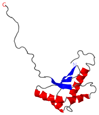
NMR StructureChain A from PDB Type:PROTEIN Length:129 aligned with RBFA_HUMAN | Q8N0V3 from UniProtKB/Swiss-Prot Length:343 Alignment length:157 88 98 108 118 128 138 148 158 168 178 188 198 208 218 228 RBFA_HUMAN 79 RSTSKKTRKEDHARLRALNGLLYKALTDLLCTPEVSQELYDLNVELSKVSLTPDFSACRAYWKTTLSAEQNAHMEAVLQRSAAHMRHLLMSQQTLRNVPPIVFVQDKGNAALAELDQLLAVADFGPRDERDNFVQNDFRDPDAPQPCGTTEPTTSSS 235 SCOP domains -------d2e7ga1 A:86-201 Ribosome-binding factor A, RbfA ---------------------------------- SCOP domains CATH domains ------------------------------------------------------------------------------------------------------------------------------------------------------------- CATH domains Pfam domains ------------------------------------------------------------------------------------------------------------------------------------------------------------- Pfam domains SAPs(SNPs) -------------------------------------------M----------------------------------------------------------------------------------------------------------------- SAPs(SNPs) PROSITE ------------------------------------------------------------------------------------RBFA PDB: A:163-184 --------------------------------------------------- PROSITE Transcript 1 (1) Exon 1.3 PDB: A:79-126 UniProt: 68-126 Exon 1.4 PDB: A:127-164 ----------------------------Exon 1.6 PDB: A:193-204 ------------------ Transcript 1 (1) Transcript 1 (2) -------------------------------------------------------------------------------------Exon 1.5 PDB: A:164-192 ------------------------Exon 1.7b Transcript 1 (2) 2e7g A 79 GSSGSSGRKEDHARLRALNGLLYKALTDLLCTPEVSQELYDLNVELSKVSLTPDFSACRAYWKTTLSAEQNAHMEAVLQRSAAHMRHLLMSQQTLRNVPPIVFVQDKGNAALAELDQLLAVADSGP----------------------------SSG 207 88 98 108 118 128 138 148 158 168 178 188 198 | - - - | 204 205
|
||||||||||||||||||||
SCOP Domains (1, 1)
NMR Structure
 |
CATH Domains (0, 0)| (no "CATH Domain" information available for 2E7G) |
Pfam Domains (0, 0)| (no "Pfam Domain" information available for 2E7G) |
Gene Ontology (6, 6)|
NMR Structure(hide GO term definitions) Chain A (RBFA_HUMAN | Q8N0V3)
|
||||||||||||||||||||||||||||||||||||||||||||||||||||||
Interactive Views
|
||||||||||||||||||||||||||||||||||||||||||||||||||||||||||||||||||||||||||||||||||||||||||||||||||||||||||||||||||||
Still Images
|
||||||||||||||||
Databases
|
||||||||||||||||||||||||||||||||||||||||||||||||||||||||||||||||||||||||||||||||||||||||||||||||||||||||||||||||||||||||||||||||||||||||||||||||||||||||||||||||
Analysis Tools
|
|||||||||||||||||||||||||||||||||||||||||||||||||||||||||||||
Entries Sharing at Least One Protein Chain (UniProt ID)
Related Entries Specified in the PDB File
|
|