|
|
|
|
 Description
|
|
Compounds
|
||||||||||||||||||||||||||||||||||||||||||||||||||||
Chains, Units
Summary Information (see also Sequences/Alignments below) |
Ligands, Modified Residues, Ions (0, 0)| (no "Ligand,Modified Residues,Ions" information available for 2CVR) |
Sites (0, 0)| (no "Site" information available for 2CVR) |
SS Bonds (0, 0)| (no "SS Bond" information available for 2CVR) |
Cis Peptide Bonds (6, 10)
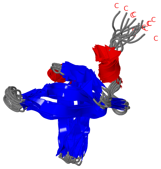
NMR Structure
|
|||||||||||||||||||||||||||||||||||
SAPs(SNPs)/Variants (0, 0)| (no "SAP(SNP)/Variant" information available for 2CVR) |
PROSITE Motifs (0, 0)| (no "PROSITE Motif" information available for 2CVR) |
Exons (0, 0)| (no "Exon" information available for 2CVR) |
Sequences/Alignments
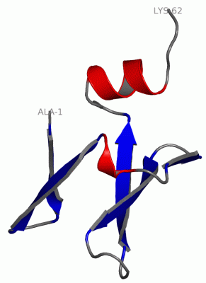
NMR StructureChain A from PDB Type:PROTEIN Length:62 aligned with DN7A_SULSO | P61991 from UniProtKB/Swiss-Prot Length:64 Alignment length:62 11 21 31 41 51 61 DN7A_SULSO 2 ATVKFKYKGEEKQVDISKIKKVWRVGKMISFTYDEGGGKTGRGAVSEKDAPKELLQMLEKQK 63 SCOP domains d2cvra_ A: DNA-binding protein SCOP domains CATH domains 2cvrA00 A:1-62 [code=2.40.50.40, no name defined] CATH domains Pfam domains -------------------------------------------------------------- Pfam domains SAPs(SNPs) -------------------------------------------------------------- SAPs(SNPs) PROSITE -------------------------------------------------------------- PROSITE Transcript -------------------------------------------------------------- Transcript 2cvr A 1 ATVKFKYKGEELQVDISKIKKVWRVGKMISFTYDEGGGKTGRGAVSEKDAPKELLQMLEKQK 62 10 20 30 40 50 60
|
||||||||||||||||||||
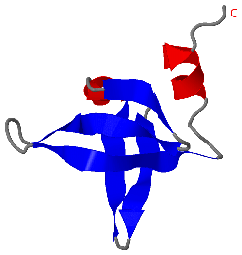
SCOP Domains (1, 1)
NMR Structure
 |
CATH Domains (1, 1)
NMR Structure
|
Pfam Domains (0, 0)| (no "Pfam Domain" information available for 2CVR) |
Gene Ontology (4, 4)|
NMR Structure(hide GO term definitions) Chain A (DN7A_SULSO | P61991)
|
||||||||||||||||||||||||||||||||||||
Interactive Views
|
||||||||||||||||||||||||||||||||||||||||||||||||||||||||||||||||||||||||||||||||||||||||||||||||||||||||||||||||||||||||||||||||||||||||||||||||||||||||
Still Images
|
||||||||||||||||
Databases
|
||||||||||||||||||||||||||||||||||||||||||||||||||||||||||||||||||||||||||||||||||||||||||||||||||||||||||||||||||||||||||||||||||||||||||||||||||||||||||||||||
Analysis Tools
|
|||||||||||||||||||||||||||||||||||||||||||||||||||||||||||||
Entries Sharing at Least One Protein Chain (UniProt ID)
Related Entries Specified in the PDB File
|
|