| molecular function |
|---|
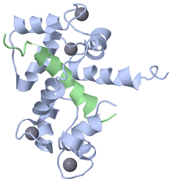
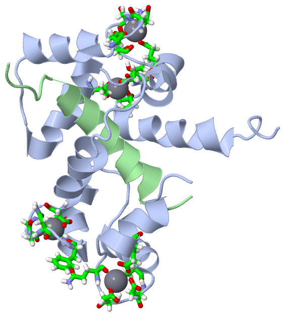
| | GO:0005509 | | calcium ion binding | | Interacting selectively and non-covalently with calcium ions (Ca2+). |
| | GO:0046872 | | metal ion binding | | Interacting selectively and non-covalently with any metal ion. |
| | GO:0031489 | | myosin V binding | | Interacting selectively and non-covalently with a class V myosin; myosin V is a dimeric molecule involved in intracellular transport. |
| | GO:0070855 | | myosin VI head/neck binding | | Interacting selectively and non-covalently with the head/neck region of a myosin VI heavy chain. |
| | GO:0032036 | | myosin heavy chain binding | | Interacting selectively and non-covalently with a heavy chain of a myosin complex. |
| | GO:0005515 | | protein binding | | Interacting selectively and non-covalently with any protein or protein complex (a complex of two or more proteins that may include other nonprotein molecules). |
| biological process |
|---|
| | GO:0030048 | | actin filament-based movement | | Movement of organelles or other particles along actin filaments, or sliding of actin filaments past each other, mediated by motor proteins. |
| | GO:0016062 | | adaptation of rhodopsin mediated signaling | | The process in which a rhodopsin-mediated signaling pathway is adjusted to modulate the sensitivity and response of a visual system to light stimuli (that might vary over more than 6 magnitudes in intensity) without response saturation. |
| | GO:0007099 | | centriole replication | | The cell cycle process in which a daughter centriole is formed perpendicular to an existing centriole. An immature centriole contains a ninefold radially symmetric array of single microtubules; mature centrioles consist of a radial array of nine microtubule triplets, doublets, or singlets depending upon the species and cell type. Duplicated centrioles also become the ciliary basal body in cells that form cilia during G0. |
| | GO:0016059 | | deactivation of rhodopsin mediated signaling | | The process of restoring the photoreceptor cell to its unexcited state after termination of the stimulus (photon). |
| | GO:0051383 | | kinetochore organization | | A process that is carried out at the cellular level which results in the assembly, arrangement of constituent parts, or disassembly of the kinetochore, a multisubunit complex that is located at the centromeric region of DNA and provides an attachment point for the spindle microtubules. |
| | GO:0046331 | | lateral inhibition | | Signaling between cells of equivalent developmental potential that results in these cells adopting different developmental fates. An example is the suppression by cells with a particular fate of the adoption of the same fate by surrounding cells. |
| | GO:0016060 | | metarhodopsin inactivation | | The process in which metarhodopsin is prevented from generating molecular signals. Activated rhodopsin (R*) is inactivated by a two-step process: first, R* is phosphorylated by rhodopsin kinase which lowers the activity of R*. Second, the protein arrestin binds to phosphorylated R* to de-activate it. |
| | GO:0090307 | | mitotic spindle assembly | | The aggregation, arrangement and bonding together of a set of components to form the spindle that contributes to the process of mitosis. |
| | GO:0007052 | | mitotic spindle organization | | A process that is carried out at the cellular level which results in the assembly, arrangement of constituent parts, or disassembly of the microtubule spindle during a mitotic cell cycle. |
| | GO:0046716 | | muscle cell cellular homeostasis | | The cellular homeostatic process that preserves a muscle cell in a stable functional or structural state. |
| | GO:0072499 | | photoreceptor cell axon guidance | | The chemotaxis process that directs the migration of a photoreceptor cell axon growth cone to its target in the optic lobe in response to a combination of attractive and repulsive cues. |
| | GO:0051533 | | positive regulation of NFAT protein import into nucleus | | Any process that activates or increases the frequency, rate or extent of the movement of an NFAT protein from the cytoplasm to the nucleus. |
| | GO:2001259 | | positive regulation of cation channel activity | | Any process that activates or increases the frequency, rate or extent of cation channel activity. |
| | GO:0006468 | | protein phosphorylation | | The process of introducing a phosphate group on to a protein. |
| | GO:0016061 | | regulation of light-activated channel activity | | Any process that modulates the frequency, rate or extent of light-activated channel activity. |
| | GO:2001020 | | regulation of response to DNA damage stimulus | | Any process that modulates the frequency, rate or extent of response to DNA damage stimulus. |
| | GO:0042052 | | rhabdomere development | | The assembly and arrangement of a rhabdomere within a cell. The rhabdomere is the organelle on the apical surface of a photoreceptor cell that contains the visual pigments. |
| | GO:0016056 | | rhodopsin mediated signaling pathway | | The series of molecular signals generated as a consequence of excitation of rhodopsin by a photon and the events that convert the absorbed photons into a cellular response. |
| | GO:0035071 | | salivary gland cell autophagic cell death | | The stage-specific programmed cell death of salivary gland cells during salivary gland histolysis. |
| | GO:0007608 | | sensory perception of smell | | The series of events required for an organism to receive an olfactory stimulus, convert it to a molecular signal, and recognize and characterize the signal. Olfaction involves the detection of chemical composition of an organism's ambient medium by chemoreceptors. This is a neurological process. |
| | GO:0007605 | | sensory perception of sound | | The series of events required for an organism to receive an auditory stimulus, convert it to a molecular signal, and recognize and characterize the signal. Sonic stimuli are detected in the form of vibrations and are processed to form a sound. |
| cellular component |
|---|
| | GO:0005814 | | centriole | | A cellular organelle, found close to the nucleus in many eukaryotic cells, consisting of a small cylinder with microtubular walls, 300-500 nm long and 150-250 nm in diameter. It contains nine short, parallel, peripheral microtubular fibrils, each fibril consisting of one complete microtubule fused to two incomplete microtubules. Cells usually have two centrioles, lying at right angles to each other. At division, each pair of centrioles generates another pair and the twin pairs form the pole of the mitotic spindle. |
| | GO:0005813 | | centrosome | | A structure comprised of a core structure (in most organisms, a pair of centrioles) and peripheral material from which a microtubule-based structure, such as a spindle apparatus, is organized. Centrosomes occur close to the nucleus during interphase in many eukaryotic cells, though in animal cells it changes continually during the cell-division cycle. |
| | GO:0005737 | | cytoplasm | | All of the contents of a cell excluding the plasma membrane and nucleus, but including other subcellular structures. |
| | GO:0005829 | | cytosol | | The part of the cytoplasm that does not contain organelles but which does contain other particulate matter, such as protein complexes. |
| | GO:0005875 | | microtubule associated complex | | Any multimeric complex connected to a microtubule. |
| | GO:0030496 | | midbody | | A thin cytoplasmic bridge formed between daughter cells at the end of cytokinesis. The midbody forms where the contractile ring constricts, and may persist for some time before finally breaking to complete cytokinesis. |
| | GO:0031475 | | myosin V complex | | A myosin complex containing a dimer of class V myosin heavy chains and associated light chains; involved in intracellular transport. Myosin V is a dimeric molecule consisting of conserved motor domains followed by 6 IQ motifs which bind specific light chains and calmodulin. The tail domain is important for cellular localization and cargo binding and can be divided into an alpha-helical coiled coil region and a C-terminal globular region. |
| | GO:0031476 | | myosin VI complex | | A myosin complex containing one or more class VI myosin heavy chains and associated light chains. Myosin VI has a single IQ motif in the neck and a tail region with a coiled coil domain followed by a unique globular domain; a unique insertion that enables myosin VI to move towards the pointed or minus end of actin filaments. |
| | GO:0005654 | | nucleoplasm | | That part of the nuclear content other than the chromosomes or the nucleolus. |
| | GO:0005886 | | plasma membrane | | The membrane surrounding a cell that separates the cell from its external environment. It consists of a phospholipid bilayer and associated proteins. |
| | GO:0016028 | | rhabdomere | | The specialized microvilli-containing organelle on the apical surfaces of a photoreceptor cell containing the visual pigment rhodopsin and most of the proteins involved in phototransduction. |
| | GO:0005819 | | spindle | | The array of microtubules and associated molecules that forms between opposite poles of a eukaryotic cell during mitosis or meiosis and serves to move the duplicated chromosomes apart. |
| | GO:0000922 | | spindle pole | | Either of the ends of a spindle, where spindle microtubules are organized; usually contains a microtubule organizing center and accessory molecules, spindle microtubules and astral microtubules. |
 Description