Show PDB file:   
         Plain Text   HTML   (compressed file size)
QuickSearch:   
by PDB,NDB,UniProt,PROSITE Code or Search Term(s)  
(-)Asymmetric Unit
(-)Asym. Unit - sites
(-)Biological Unit 1
(-)Biol. Unit 1 - sites
collapse expand < >
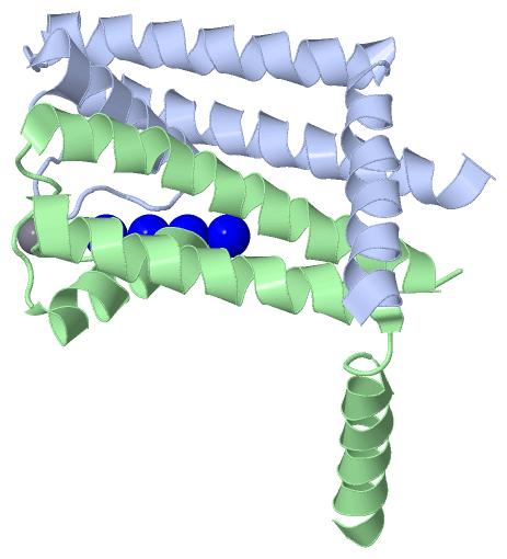
Image Asymmetric Unit
Asymmetric Unit  (Jmol Viewer)
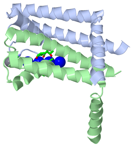
Image Asym. Unit - sites
Asym. Unit - sites  (Jmol Viewer)
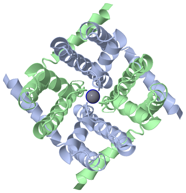
Image Biological Unit 1
Biological Unit 1  (Jmol Viewer)
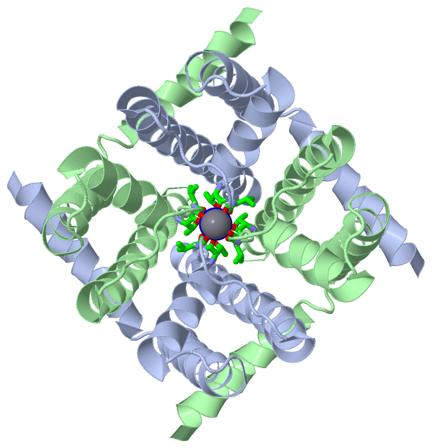
Image Biol. Unit 1 - sites
Biol. Unit 1 - sites  (Jmol Viewer)

(-) Description

Title :  NA+ COMPLEX OF THE NAK CHANNEL
 
Authors :  N. Shi, S. Ye, A. Alam, L. Chen, Y. Jiang
Date :  28 Jul 05  (Deposition) - 07 Feb 06  (Release) - 13 Jul 11  (Revision)
Method :  X-RAY DIFFRACTION
Resolution :  2.40
Chains :  Asym. Unit :  A,B
Biol. Unit 1:  A,B  (2x)
Keywords :  Inverted Teepee, Helix Bundle, Tetramer, Central Cavity, Ion Binding, Transport Protein (Keyword Search: [Gene Ontology, PubMed, Web (Google))
 
Reference :  N. Shi, S. Ye, A. Alam, L. Chen, Y. Jiang
Atomic Structure Of A Na+- And K+-Conducting Channel.
Nature V. 440 570 2006
PubMed-ID: 16467789  |  Reference-DOI: 10.1038/NATURE04508

(-) Compounds

Molecule 1 - POTASSIUM CHANNEL PROTEIN
    ChainsA, B
    EngineeredYES
    Expression SystemESCHERICHIA COLI
    Expression System PlasmidPQE 60
    Expression System StrainXL-1 BLUE
    Expression System Taxid562
    Expression System Vector TypePLASMID
    Organism ScientificBACILLUS CEREUS
    Organism Taxid1396

 Structural Features

(-) Chains, Units

  12
Asymmetric Unit AB
Biological Unit 1 (2x)AB

Summary Information (see also Sequences/Alignments below)

(-) Ligands, Modified Residues, Ions  (2, 5)

Asymmetric Unit (2, 5)
No.NameCountTypeFull Name
1CA1Ligand/IonCALCIUM ION
2NA4Ligand/IonSODIUM ION
Biological Unit 1 (0, 0)
No.NameCountTypeFull Name
1CA-1Ligand/IonCALCIUM ION
2NA-1Ligand/IonSODIUM ION

(-) Sites  (3, 3)

Asymmetric Unit (3, 3)
No.NameEvidenceResiduesDescription
1AC1SOFTWARETHR A:63 , VAL A:64 , THR B:63 , VAL B:64BINDING SITE FOR RESIDUE NA A 111
2AC2SOFTWARETHR A:63 , THR B:63BINDING SITE FOR RESIDUE NA A 112
3AC3SOFTWAREGLY A:67 , GLY B:67BINDING SITE FOR RESIDUE CA A 114

(-) SS Bonds  (0, 0)

(no "SS Bond" information available for 2AHY)

(-) Cis Peptide Bonds  (0, 0)

(no "Cis Peptide Bond" information available for 2AHY)

 Sequence-Structure Mapping

(-) SAPs(SNPs)/Variants  (0, 0)

(no "SAP(SNP)/Variant" information available for 2AHY)

(-) PROSITE Motifs  (0, 0)

(no "PROSITE Motif" information available for 2AHY)

(-) Exons   (0, 0)

(no "Exon" information available for 2AHY)

(-) Sequences/Alignments

Asymmetric Unit
   Reformat: Number of residues per line =  ('0' or empty: single-line sequence representation)
  Number of residues per labelling interval =   
  UniProt sequence: complete  aligned part    
   Show mapping: SCOP domains CATH domains Pfam domains Secondary structure (by author)
SAPs(SNPs) PROSITE motifs Exons
(details for a mapped element are shown in a popup box when the mouse pointer rests over it)
Chain A from PDB  Type:PROTEIN  Length:104
 aligned with Q81HW2_BACCR | Q81HW2 from UniProtKB/TrEMBL  Length:114

    Alignment length:104
                                    10        20        30        40        50        60        70        80        90       100    
         Q81HW2_BACCR     1 MLSFLLTLKRMLRACLRAWKDKEFQVLFVLTILTLISGTIFYSTVEGLRPIDALYFSVVTLTTVGDGNFSPQTDFGKIFTILYIFIGIGLVFGFIHKLAVNVQL 104
               SCOP domains -------------------------------------------------------------------------------------------------------- SCOP domains
               CATH domains 2ahyA00 A:1-104  [code=1.10.287.70, no name defined]                                                     CATH domains
               Pfam domains -------------------------------------------------------------------------------------------------------- Pfam domains
         Sec.struct. author hhhhhhhhhhhhhhhhhhh.hhhhhhhhhhhhhhhhhhhhhhhhh...hhhhhhhhhhhhhh..........hhhhhhhhhhhhhhhhhhhhhhhhhhhhhhhh Sec.struct. author
                 SAPs(SNPs) -------------------------------------------------------------------------------------------------------- SAPs(SNPs)
                    PROSITE -------------------------------------------------------------------------------------------------------- PROSITE
                 Transcript -------------------------------------------------------------------------------------------------------- Transcript
                 2ahy A   1 MLSFLLTLKRMLRACLRAWKDKEFQVLFVLTILTLISGTIFYSTVEGLRPIDALYFSVVTLTTVGDGNFSPQTDFGKIFTILYIFIGIGLVFGFIHKLAVNVQL 104
                                    10        20        30        40        50        60        70        80        90       100    

Chain B from PDB  Type:PROTEIN  Length:103
 aligned with Q81HW2_BACCR | Q81HW2 from UniProtKB/TrEMBL  Length:114

    Alignment length:103
                                    10        20        30        40        50        60        70        80        90       100   
         Q81HW2_BACCR     1 MLSFLLTLKRMLRACLRAWKDKEFQVLFVLTILTLISGTIFYSTVEGLRPIDALYFSVVTLTTVGDGNFSPQTDFGKIFTILYIFIGIGLVFGFIHKLAVNVQ 103
               SCOP domains ------------------------------------------------------------------------------------------------------- SCOP domains
               CATH domains 2ahyB00 B:1-103  [code=1.10.287.70, no name defined]                                                    CATH domains
               Pfam domains ------------------------------------------------------------------------------------------------------- Pfam domains
         Sec.struct. author hhhhhhhhhhhhhhhhh...hhhhhhhhhhhhhhhhhhhhhhhhh...hhhhhhhhhhhhhh..........hhhhhhhhhhhhhhhhhhhhhhhhhhhhh.. Sec.struct. author
                 SAPs(SNPs) ------------------------------------------------------------------------------------------------------- SAPs(SNPs)
                    PROSITE ------------------------------------------------------------------------------------------------------- PROSITE
                 Transcript ------------------------------------------------------------------------------------------------------- Transcript
                 2ahy B   1 MLSFLLTLKRMLRACLRAWKDKEFQVLFVLTILTLISGTIFYSTVEGLRPIDALYFSVVTLTTVGDGNFSPQTDFGKIFTILYIFIGIGLVFGFIHKLAVNVQ 103
                                    10        20        30        40        50        60        70        80        90       100   

   Legend:   → Mismatch (orange background)
  - → Gap (green background, '-', border residues have a numbering label)
    → Modified Residue (blue background, lower-case, 'x' indicates undefined single-letter code, labelled with number + name)
  x → Chemical Group (purple background, 'x', labelled with number + name, e.g. ACE or NH2)
  extra numbering lines below/above indicate numbering irregularities and modified residue names etc., number ends below/above '|'

 Classification and Annotation

(-) SCOP Domains  (0, 0)

(no "SCOP Domain" information available for 2AHY)

(-) CATH Domains  (1, 2)

Asymmetric Unit

(-) Pfam Domains  (0, 0)

(no "Pfam Domain" information available for 2AHY)

(-) Gene Ontology  (2, 2)

Asymmetric Unit(hide GO term definitions)
Chain A,B   (Q81HW2_BACCR | Q81HW2)
cellular component
    GO:0016021    integral component of membrane    The component of a membrane consisting of the gene products and protein complexes having at least some part of their peptide sequence embedded in the hydrophobic region of the membrane.
    GO:0016020    membrane    A lipid bilayer along with all the proteins and protein complexes embedded in it an attached to it.

 Visualization

(-) Interactive Views

Asymmetric Unit
  Complete Structure
    Jena3D(integrated viewing of ligand, site, SAP, PROSITE, SCOP information)
    WebMol | AstexViewer[tm]@PDBe
(Java Applets, require no local installation except for Java; loading may be slow)
    STRAP
(Java WebStart application, automatic local installation, requires Java; full application with system access!)
    RasMol
(require local installation)
    Molscript (VRML)
(requires installation of a VRML viewer; select preferred view via VRML and generate a mono or stereo PDF format file)
 
  Ligands, Modified Residues, Ions
    CA  [ RasMol | Jena3D ]  +environment [ RasMol | Jena3D ]
    NA  [ RasMol | Jena3D ]  +environment [ RasMol | Jena3D ]
 
  Sites
    AC1  [ RasMol ]  +environment [ RasMol ]
    AC2  [ RasMol ]  +environment [ RasMol ]
    AC3  [ RasMol ]  +environment [ RasMol ]
 
  Cis Peptide Bonds
(no "Cis Peptide Bonds" information available for 2ahy)
 
Biological Unit
  Complete Structure
    Biological Unit 1  [ Jena3D ]

(-) Still Images

Jmol
  protein: cartoon or spacefill or dots and stick; nucleic acid: cartoon and stick; ligands: spacefill; active site: stick
Molscript
  protein, nucleic acid: cartoon; ligands: spacefill; active site: ball and stick

 Databases and Analysis Tools

(-) Databases

Access by PDB/NDB ID
  2ahy
    Family and Domain InformationProDom | SYSTERS
    General Structural InformationGlycoscienceDB | MMDB | NDB | OCA | PDB | PDBe | PDBj | PDBsum | PDBWiki | PQS | PROTEOPEDIA
    Orientation in MembranesOPM
    Protein SurfaceSURFACE
    Secondary StructureDSSP (structure derived) | HSSP (homology derived)
    Structural GenomicsGeneCensus
    Structural NeighboursCE | VAST
    Structure ClassificationCATH | Dali | SCOP
    Validation and Original DataBMRB Data View | BMRB Restraints Grid | EDS | PROCHECK | RECOORD | WHAT_CHECK
 
Access by UniProt ID/Accession number
  Q81HW2_BACCR | Q81HW2
    Comparative Protein Structure ModelsModBase
    Genomic InformationEnsembl
    Protein-protein InteractionDIP
    Sequence, Family and Domain InformationInterPro | Pfam | SMART | UniProtKB/TrEMBL
 
Access by Enzyme Classificator   (EC Number)
  (no 'Enzyme Classificator' available)
    General Enzyme InformationBRENDA | EC-PDB | Enzyme | IntEnz
    PathwayKEGG | MetaCyc
 
Access by Disease Identifier   (MIM ID)
  (no 'MIM ID' available)
    Disease InformationOMIM
 
Access by GenAge ID
  (no 'GenAge ID' available)
    Age Related InformationGenAge

(-) Analysis Tools

Access by PDB/NDB ID
    Domain InformationXDom
    Interatomic Contacts of Structural UnitsCSU
    Ligand-protein ContactsLPC
    Protein CavitiescastP
    Sequence and Secondary StructurePDBCartoon
    Structure AlignmentSTRAP(Java WebStart application, automatic local installation, requires Java; full application with system access!)
    Structure and Sequence BrowserSTING
 
Access by UniProt ID/Accession number
  Q81HW2_BACCR | Q81HW2
    Protein Disorder PredictionDisEMBL | FoldIndex | GLOBPLOT (for more information see DisProt)

 Related Entries

(-) Entries Sharing at Least One Protein Chain (UniProt ID)

UniProtKB/TrEMBL
        Q81HW2_BACCR | Q81HW22ahz 2q67 2q68 2q69 2q6a 3e83 3e86 3e89 3e8b 3e8f 3e8g 3e8h 3k03 3k04 3k06 3k08 3k0d 3k0g 3t1c 3t2m 3t4d 3t4z 3tcu 3tet 4pdl 4pdm 4pdr 4pdv 4r50 4r6z 4r7c 4r8c 4rai 4ro2 4zbm

(-) Related Entries Specified in the PDB File

2ahz K+ COMPLEX OF THE NAK CHANNEL