| molecular function |
|---|
| | GO:0043754 | | dihydrolipoyllysine-residue (2-methylpropanoyl)transferase activity | | Catalysis of the reaction: 2-methylpropanoyl-CoA + enzyme N6-(dihydrolipoyl)lysine = CoA + enzyme N6-(S-[2-methylpropanoyl]dihydrolipoyl)lysine. |
| | GO:0016740 | | transferase activity | | Catalysis of the transfer of a group, e.g. a methyl group, glycosyl group, acyl group, phosphorus-containing, or other groups, from one compound (generally regarded as the donor) to another compound (generally regarded as the acceptor). Transferase is the systematic name for any enzyme of EC class 2. |
| | GO:0016746 | | transferase activity, transferring acyl groups | | Catalysis of the transfer of an acyl group from one compound (donor) to another (acceptor). |
| | GO:0031625 | | ubiquitin protein ligase binding | | Interacting selectively and non-covalently with a ubiquitin protein ligase enzyme, any of the E3 proteins. |
| biological process |
|---|
| | GO:0009083 | | branched-chain amino acid catabolic process | | The chemical reactions and pathways resulting in the breakdown of amino acids containing a branched carbon skeleton, comprising isoleucine, leucine and valine. |
| | GO:0046487 | | glyoxylate metabolic process | | The chemical reactions and pathways involving glyoxylate, the anion of glyoxylic acid, HOC-COOH. |
| | GO:0008152 | | metabolic process | | The chemical reactions and pathways, including anabolism and catabolism, by which living organisms transform chemical substances. Metabolic processes typically transform small molecules, but also include macromolecular processes such as DNA repair and replication, and protein synthesis and degradation. |
| cellular component |
|---|
| | GO:0005947 | | mitochondrial alpha-ketoglutarate dehydrogenase complex | | Mitochondrial complex that possesses alpha-ketoglutarate dehydrogenase activity. |
| | GO:0005759 | | mitochondrial matrix | | The gel-like material, with considerable fine structure, that lies in the matrix space, or lumen, of a mitochondrion. It contains the enzymes of the tricarboxylic acid cycle and, in some organisms, the enzymes concerned with fatty acid oxidation. |
| | GO:0042645 | | mitochondrial nucleoid | | The region of a mitochondrion to which the DNA is confined. |
| | GO:0005739 | | mitochondrion | | A semiautonomous, self replicating organelle that occurs in varying numbers, shapes, and sizes in the cytoplasm of virtually all eukaryotic cells. It is notably the site of tissue respiration. |
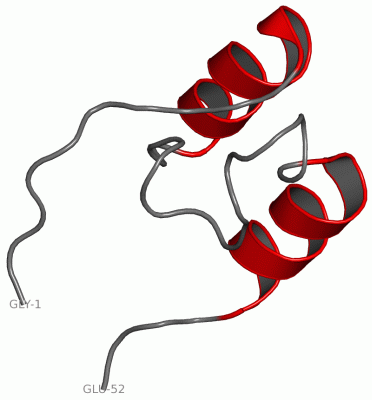
 Description
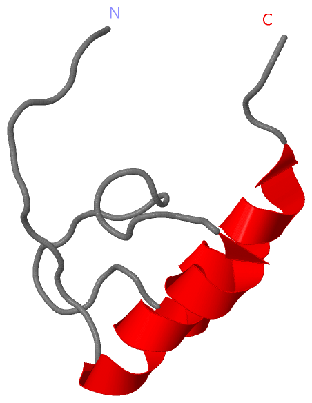