|
|
|
|
 Description
|
|
Compounds
|
||||||||||||||||||||||||||||||||||||||||||||||||
Chains, Units
Summary Information (see also Sequences/Alignments below) |
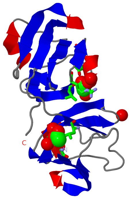
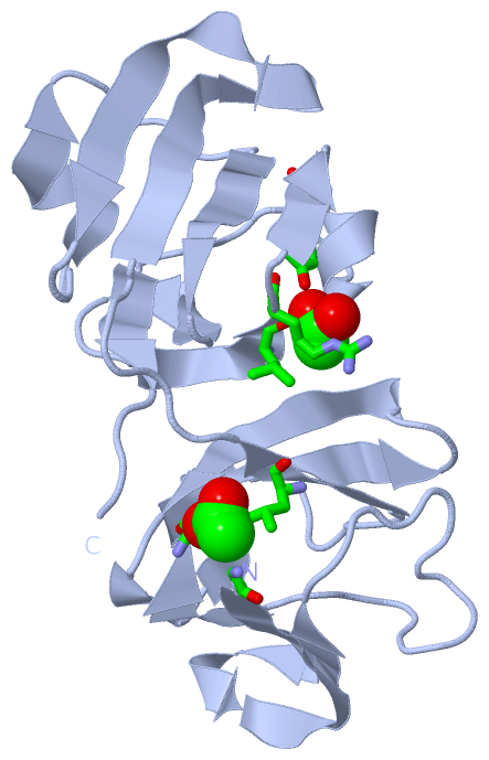
Ligands, Modified Residues, Ions (2, 204)| Asymmetric Unit (2, 204) Biological Unit 1 (1, 2) |
Sites (2, 2)
Asymmetric Unit (2, 2)
|
SS Bonds (0, 0)| (no "SS Bond" information available for 1ZIE) |
Cis Peptide Bonds (0, 0)| (no "Cis Peptide Bond" information available for 1ZIE) |
SAPs(SNPs)/Variants (0, 0)| (no "SAP(SNP)/Variant" information available for 1ZIE) |
PROSITE Motifs (1, 4)
Asymmetric Unit (1, 4)
|
||||||||||||||||||||||||||||||||||||||||||||||||
Exons (0, 0)| (no "Exon" information available for 1ZIE) |
Sequences/Alignments
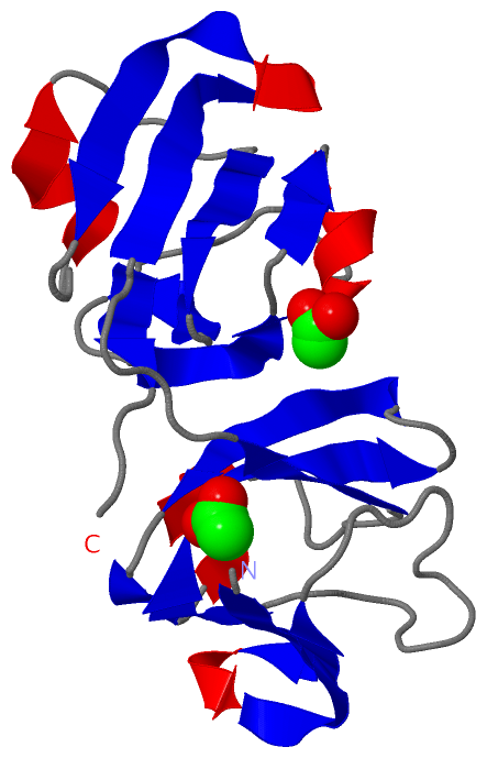
Asymmetric UnitChain A from PDB Type:PROTEIN Length:172 aligned with CRGE_RAT | P02528 from UniProtKB/Swiss-Prot Length:174 Alignment length:172 11 21 31 41 51 61 71 81 91 101 111 121 131 141 151 161 171 CRGE_RAT 2 GKITFYEDRGFQGRHYECSTDHSNLQPYFSRCNSVRVDSGCWMLYEQPNFTGCQYFLRRGDYPDYQQWMGFSDSVRSCRLIPHSSSHRIRIYEREDYRGQMVEITDDCPHLQDRFHFSDFHSFHVMEGYWVLYEMPNYRGRQYLLRPGEYRRYHDWGAMNARVGSLRRIMDF 173 SCOP domains d1ziea1 A:1001-1084 beta-Crystallin d1ziea2 A:1085-1172 beta-Crystallin SCOP domains CATH domains 1zieA01 A:1001-1083 Crystallins 1zieA02 A:1084-1172 Crystallins CATH domains Pfam domains (1) ---------------------------------------------------------------------------------------Crystall-1zieA01 A:1088-1169 --- Pfam domains (1) Pfam domains (2) ---------------------------------------------------------------------------------------Crystall-1zieA02 A:1088-1169 --- Pfam domains (2) SAPs(SNPs) ---------------------------------------------------------------------------------------------------------------------------------------------------------------------------- SAPs(SNPs) PROSITE CRYSTALLIN_BETA_GAMMA PDB: A:1001-1039CRYSTALLIN_BETA_GAMMA PDB: A:1040-1082 ----CRYSTALLIN_BETA_GAMMA PDB: A:1087-1127 CRYSTALLIN_BETA_GAMMA PDB: A:1128-1170 -- PROSITE Transcript ---------------------------------------------------------------------------------------------------------------------------------------------------------------------------- Transcript 1zie A 1001 GKITFYEDRGFQGRHYECSTDHSNLQPYFSRCNSVRVDSGCWMLYEQPNFTGCQYFLRRGDYPDYQQWMGFSDSVRSCRLIPHSSSHRIRIYEREDYRGQMVEITDDCPHLQDRFHFSDFHSFHVMEGYWVLYEMPNYRGRQYLLRPGEYRRYHDWGAMNARVGSLRRIMDF 1172 1010 1020 1030 1040 1050 1060 1070 1080 1090 1100 1110 1120 1130 1140 1150 1160 1170
|
||||||||||||||||||||
SCOP Domains (1, 2)
Asymmetric Unit
 |
CATH Domains (1, 2)
Asymmetric Unit
|
Pfam Domains (1, 2)
Asymmetric Unit
|
Gene Ontology (2, 2)|
Asymmetric Unit(hide GO term definitions) Chain A (CRGE_RAT | P02528)
|
||||||||||||||||||||||||
Interactive Views
|
||||||||||||||||||||||||||||||||||||||||||||||||||||||||||||||||||||||||||||||||||||||||||||||||||||||||||||||||||||||||||||||||||||||||||||||||||||||
Still Images
|
||||||||||||||||
Databases
|
||||||||||||||||||||||||||||||||||||||||||||||||||||||||||||||||||||||||||||||||||||||||||||||||||||||||||||||||||||||||||||||||||||||||||||||||||||||||||||||||
Analysis Tools
|
|||||||||||||||||||||||||||||||||||||||||||||||||||||||||||||
Entries Sharing at Least One Protein Chain (UniProt ID)
Related Entries Specified in the PDB File
|
|