| molecular function |
|---|
| | GO:0003993 | | acid phosphatase activity | | Catalysis of the reaction: an orthophosphoric monoester + H2O = an alcohol + phosphate, with an acid pH optimum. |
| | GO:0016787 | | hydrolase activity | | Catalysis of the hydrolysis of various bonds, e.g. C-O, C-N, C-C, phosphoric anhydride bonds, etc. Hydrolase is the systematic name for any enzyme of EC class 3. |
| | GO:0004726 | | non-membrane spanning protein tyrosine phosphatase activity | | Catalysis of the reaction: non-membrane spanning protein tyrosine phosphate + H2O = non-membrane spanning protein tyrosine + phosphate. |
| | GO:0016791 | | phosphatase activity | | Catalysis of the hydrolysis of phosphoric monoesters, releasing inorganic phosphate. |
| | GO:0004721 | | phosphoprotein phosphatase activity | | Catalysis of the reaction: a phosphoprotein + H2O = a protein + phosphate. Together with protein kinases, these enzymes control the state of phosphorylation of cell proteins and thereby provide an important mechanism for regulating cellular activity. |
| | GO:0005515 | | protein binding | | Interacting selectively and non-covalently with any protein or protein complex (a complex of two or more proteins that may include other nonprotein molecules). |
| | GO:0004725 | | protein tyrosine phosphatase activity | | Catalysis of the reaction: protein tyrosine phosphate + H2O = protein tyrosine + phosphate. |
| biological process |
|---|
| | GO:0035335 | | peptidyl-tyrosine dephosphorylation | | The removal of phosphoric residues from peptidyl-O-phospho-tyrosine to form peptidyl-tyrosine. |
| | GO:0006470 | | protein dephosphorylation | | The process of removing one or more phosphoric residues from a protein. |
| cellular component |
|---|
| | GO:0005737 | | cytoplasm | | All of the contents of a cell excluding the plasma membrane and nucleus, but including other subcellular structures. |
| | GO:0009898 | | cytoplasmic side of plasma membrane | | The leaflet the plasma membrane that faces the cytoplasm and any proteins embedded or anchored in it or attached to its surface. |
| | GO:0070062 | | extracellular exosome | | A vesicle that is released into the extracellular region by fusion of the limiting endosomal membrane of a multivesicular body with the plasma membrane. Extracellular exosomes, also simply called exosomes, have a diameter of about 40-100 nm. |
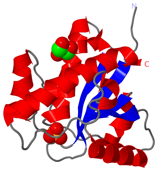
 Description
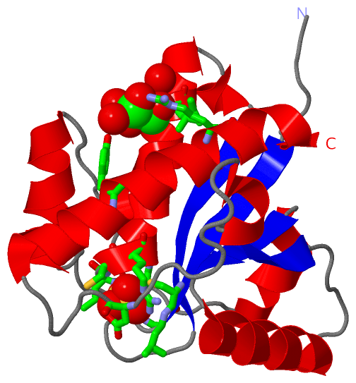