|
|
|
|
 Description
|
|
Compounds
|
||||||||||||||||||||||||||||
Chains, Units
Summary Information (see also Sequences/Alignments below) |
Ligands, Modified Residues, Ions (3, 9)
Asymmetric Unit (3, 9)
|
Sites (9, 9)
Asymmetric Unit (9, 9)
|
SS Bonds (0, 0)| (no "SS Bond" information available for 1XMX) |
Cis Peptide Bonds (1, 1)
Asymmetric Unit
|
||||||||
SAPs(SNPs)/Variants (0, 0)| (no "SAP(SNP)/Variant" information available for 1XMX) |
PROSITE Motifs (0, 0)| (no "PROSITE Motif" information available for 1XMX) |
Exons (0, 0)| (no "Exon" information available for 1XMX) |
Sequences/Alignments
Asymmetric UnitChain A from PDB Type:PROTEIN Length:380 aligned with Q9KQU9_VIBCH | Q9KQU9 from UniProtKB/TrEMBL Length:383 Alignment length:385 1 | 8 18 28 38 48 58 68 78 88 98 108 118 128 138 148 158 168 178 188 198 208 218 228 238 248 258 268 278 288 298 308 318 328 338 348 358 368 378 Q9KQU9_VIBCH - --MAIHVGIIDQDPVRLVTPLLDHRTVSRHIIFIGDHTQTVIYQRLSDVLNKRNISTDFFEIPAGSNTSAIKSAIRELAETLKARGEEVKFNASCGLRHRLLSAYEVFRSYHWPIFVVEPNSDCLCWLYPEGNNDTQVQDRITIADYLTIFGARGEFNEHQLSPQLDQQLYQLGERWASNALELGPGLATLNYLATTCRKEQKLDVELSDKQQGYRELNLLLSDLVEAKIASYENGILTFINEEARRFANGEWLETLVHSTVKQIQDDMPTIQDRSLNVQVYRQLGEREVRNELDVATVVNNKLHIIECKTKGMRDDGDDTLYKLESLRDLLGGLQARAMLVSFRPLRHNDITRAEDLGLALIGPDELKDLKTHLTQWFKAAGGN 383 SCOP domains d1xmxa_ A: Hypothetical protein VC1899 SCOP domains CATH domains ----------------------------------------------------------------------------------------------------------------------------------------------1xmxA02 -----------------------------------------------------------------------------------------1xmxA02 A:141-161,A:251-383 [code=3.40.1350.10, no name define d] CATH domains Pfam domains --DUF1887-1xmxA01 A:1-380 --- Pfam domains SAPs(SNPs) ------------------------------------------------------------------------------------------------------------------------------------------------------------------------------------------------------------------------------------------------------------------------------------------------------------------------------------------------------------------------------------------------- SAPs(SNPs) PROSITE ------------------------------------------------------------------------------------------------------------------------------------------------------------------------------------------------------------------------------------------------------------------------------------------------------------------------------------------------------------------------------------------------- PROSITE Transcript ------------------------------------------------------------------------------------------------------------------------------------------------------------------------------------------------------------------------------------------------------------------------------------------------------------------------------------------------------------------------------------------------- Transcript 1xmx A -1 NAMAIHVGIIDQDPVRLVTPLLDHRTVSRHIIFIGDHTQTVIYQRLSDVLNKRNISTDFFEIPAGSNTSAIKSAIRELAETLKARGEEVKFNASCGLRHRLLSAYEVFRSYHWPIFVVEPNSDCLCWLYPEGNNDTQVQDRITIADYLTIFGARGEFN----SPQLDQQLYQLGERWASNALELGPGLATLNYLATTCRKEQKLDVELSDKQQGYRELNLLLSDLVEAKIASYENGILTFINEEARRFANGEWLETLVHSTVKQIQDDMPTIQDRSLNVQVYRQLGEREVRNELDVATVVNNKLHIIECKTKGMR-DGDDTLYKLESLRDLLGGLQARAMLVSFRPLRHNDITRAEDLGLALIGPDELKDLKTHLTQWFKAAGGN 383 8 18 28 38 48 58 68 78 88 98 108 118 128 138 148 | - | 168 178 188 198 208 218 228 238 248 258 268 278 288 298 308 | |318 328 338 348 358 368 378 156 161 313 | 315
|
||||||||||||||||||||
SCOP Domains (1, 1)
Asymmetric Unit
 |
CATH Domains (1, 1)
Asymmetric Unit
|
Pfam Domains (1, 1)
Asymmetric Unit
|
Gene Ontology (6, 6)|
Asymmetric Unit(hide GO term definitions) Chain A (Q9KQU9_VIBCH | Q9KQU9)
|
||||||||||||||||||||||||||||||||||||||||||||||||||||||
Interactive Views
|
||||||||||||||||||||||||||||||||||||||||||||||||||||||||||||||||||||||||||||||||||||||||||||||||||||||||||||||||||||||||||||||||||||||||||||||||||||||||||||||||||||||||||||||||||||||||||||||||||||||||||||||||||||
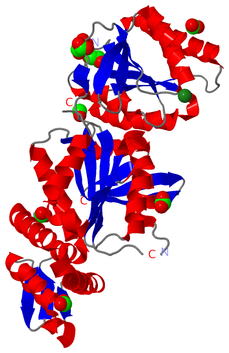
Still Images
|
||||||||||||||||
Databases
|
||||||||||||||||||||||||||||||||||||||||||||||||||||||||||||||||||||||||||||||||||||||||||||||||||||||||||||||||||||||||||||||||||||||||||||||||||||||||||||||||
Analysis Tools
|
|||||||||||||||||||||||||||||||||||||||||||||||||||||||||||||
Entries Sharing at Least One Protein Chain (UniProt ID)
Related Entries Specified in the PDB File
|
|